Team:NYMU-Taipei/ymivenusianmm.html
From 2012.igem.org
| (2 intermediate revisions not shown) | |||
| Line 63: | Line 63: | ||
<div id="ymi_header"> | <div id="ymi_header"> | ||
<div id="inner_header"> | <div id="inner_header"> | ||
| - | <a href="https://2012.igem.org/Team:NYMU-Taipei"><img src="https://static.igem.org/mediawiki/2012/ | + | <a href="https://2012.igem.org/Team:NYMU-Taipei"><img src="https://static.igem.org/mediawiki/2012/1/15/Ymi_header1.jpg" border="0"></a> |
</div> | </div> | ||
</div> | </div> | ||
| Line 150: | Line 150: | ||
<li><a title="Abstract" href="https://2012.igem.org/Team:NYMU-Taipei/ymiq1.html">Abstract</a></li> | <li><a title="Abstract" href="https://2012.igem.org/Team:NYMU-Taipei/ymiq1.html">Abstract</a></li> | ||
<li><a title="Methods" href="https://2012.igem.org/Team:NYMU-Taipei/ymiq2.html">Methods</a></li> | <li><a title="Methods" href="https://2012.igem.org/Team:NYMU-Taipei/ymiq2.html">Methods</a></li> | ||
| - | <li><a title=" | + | <li><a title="Experiments" href="https://2012.igem.org/Team:NYMU-Taipei/ymiq3.html">Experiments</a></li> |
| - | <li><a title="Results & References" href="https://2012.igem.org/Team:NYMU-Taipei/ymiq4.html">Results & References</a></li> | + | <li><a title="Results & References" href="https://2012.igem.org/Team:NYMU-Taipei/ymiq4.html">Results & References</a></li><li><a title="Further Experiments after Asia Jamboree" href="https://2012.igem.org/Team:NYMU-Taipei/ymiq5.html">Further Experiments after Asia Jamboree</a></li> |
</ul> | </ul> | ||
</li> | </li> | ||
Latest revision as of 01:51, 27 October 2012
What’s the specialty of our cyanobacteria?
Invasin(protein names: P11922 INVA_YERPS) is a protein that allows enteric bacteria to penetrate cultured mammalian cells. Listeriolysin O (LLO) facilitates bacterial escape from the internalization vesicle into the cytoplasm, where bacteria divide and undergo cell-to-cell spreading via actin-based motility[2]. And Synechococcus elongatus PCC 7942 was the first cyanobacterial strain which could be transformed by exogenously added DNA. When engineered with invasin(inv) from Yersiniapestis and listeriolysin O(llo) from Listeria monocytogenes (both provided by Pamela A. Silver, Harvard Medical school[3]), new S. elongatus was able to invade cultured mammalian cells and was capable of symbiosis with eukaryotic cells.
Additionally, invasin(inv) from Yersiniapestis and listeriolysin O(llo) from Listeria monocytogenes have been studied by 2010 Warsaw igem team. Our further study and novel observation relative to their research were posted not only on our wiki, but also on the partregistry website(BBa_K299810, BBa_K299811,BBa_K299812)
What’s the platform of coculturing?
Three types of mammalian cells were chosen—two stages of induced pluripotent stem cells from mice and J774 mouse macrophage cell line. Here are the protocols of cell culturing (two stages of mice induced pluripotent stem cells: iPS cells separated from embryonic fibroblasts (MEFs) click here, embryoid body (EB) stage of iPS cells click here, J774 mouse macrophage cell line click here)
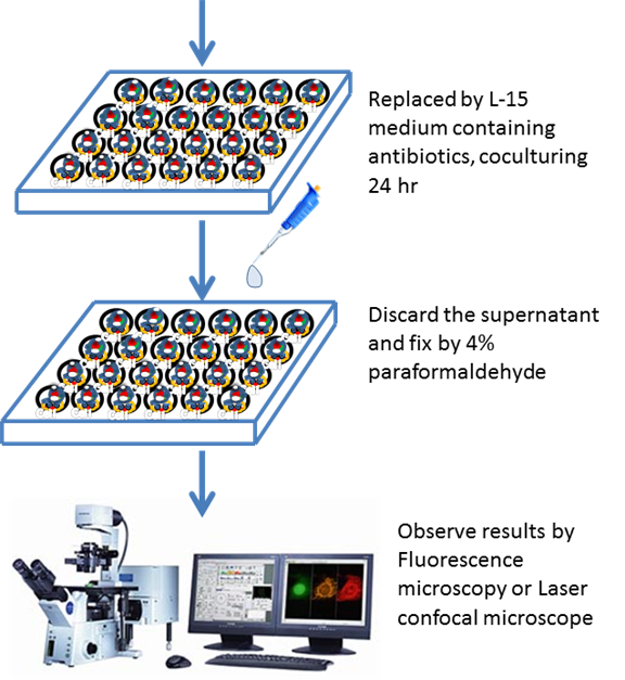
Cells were seeded on the 24-well plate. After 80% full in each well, PCC7942(wt/inv-llo transform) suspensions in PBS were set to the same OD and 50 µl of this suspension were added per 1 ml of Leibovitz's L-15 medium without phenol red, without antibiotics and with 10% fetal bovine serum (Invitrogen) per well of 24-well tissue culture dishes containing the mammalian cells. After 24-hour cells and PCC7942 coculturing under 30°C atmospheric CO2 level, supernatants were discarded and cells were washed with PBS two times and the medium was replaced by L-15 containing antibiotics, gentamycin 100ug/ml(Invitrogen). After 24 hours, cells were fixed by 4% paraformaldehyde and were observed by fluorescent microscope or confocal laser microscope.


-
Becoming Venusian
-
Sulfur Oxide Terminator
-
Sulfide as Energy Generator
-
Denitrifying Machine
-
Cd+2 Collector
 "
"
