Team:Calgary/Project/OSCAR/CatecholDegradation
From 2012.igem.org


Hello! iGEM Calgary's wiki functions best with Javascript enabled, especially for mobile devices. We recommend that you enable Javascript on your device for the best wiki-viewing experience. Thanks!
Catechol Degradation
****This section needs work. Why are we degrading catechol? What part did we use? What is the number?
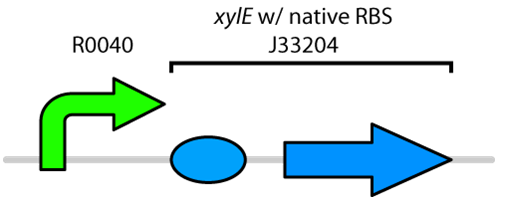
Catechol is a toxic compound found in tailings ponds that is a by-product of polyaromatic hydrocarbon metabolism. Catechol is toxic to a wide range of organisms from microorganisms to mammals (Schweigert et al., 2001). The chemical properties of catechol allow it to react with biomolecules like DNA, proteins and membranes (Schweigert et al., 2001). These interactions can cause serious damage including DNA breakage, enzyme inactivation and membrane uncoupling (Schweigert et al., 2001). Catechol can be degraded by the enzyme catechol 2,3-dioxygenase encoded by the xylE gene on the Tol plasmid of Pseudomonas putida (Nakai et al., 1983). The current iGEM Part repository has two BioBricks available of xylE. One contained xylE with its native ribosome-binding site (part: J33204), while the other part had the glucose-repressible Pcst promoter (Part: K118021). Given that E. coli is grown in the presence of glucose, we designed a new construct to keep xylE repressed by using the TetR promoter.
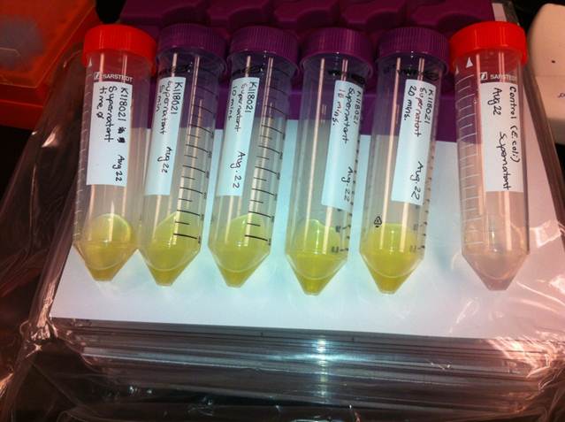
Catechol 2,3-dioxygenase is an extradiol dioxygenase which cleaves catechol adjacent to the two hydroxyl groups. When this occurs 2-hydroxymuconic semialdehyde is produced, which is yellow in colour. This change in colour allows for visual assay to assess the activity of XlyE.
The colour change assays were performed with the newly constructed part by bringing the supernatant of an overnight culture to a concentration of 0.1 M of catechol. The catechol was added to the supernatant because the reaction takes place outside of the cell. Within minutes of the addition of catechol to the supernatant, the solution turned from the pale yellow of LB to a bright yellow. This assay was completed by following the previous assay done by the 2008 Edinburgh iGEM team.
References
Nakai, C., Kagamiyama, H., and Nozaki, M. 1983. Complete nucleotide sequence of the metapyrocatechase gene on the TOL plasmid of Pseudomonas putida. The Journal of Biological Chemistry 258: 2923-2928.
Schweigert, N., Zehnder, A.J., and Eggen, R.I. 2001. Chemical properties of catechols and their molecular modes of toxic action in cells, from microorganisms to mammals. Environmental Microbiology 3: 81-91
Shu, L., Chiou, Y., Orville, A.M., Miller, M.A., Lipscomb, J.D., and Que, L. 1995. X-ray absorption spectroscopic studies of the Fe(II) active site of catechol 2,3-dioxygenase. Implications for the extradiol cleavage mechanism. Biochemistry 34: 6649-6659.
 "
"


