Team:University College London/LabBook/Week10
From 2012.igem.org
Sednanalien (Talk | contribs) (→9-6) |
Sednanalien (Talk | contribs) (→9-6) |
||
| Line 116: | Line 116: | ||
<img id="9-6" src="https://static.igem.org/mediawiki/2012/e/e6/Ucl2012-labbook-graph109-6.png" /><div class="experimentContent"> | <img id="9-6" src="https://static.igem.org/mediawiki/2012/e/e6/Ucl2012-labbook-graph109-6.png" /><div class="experimentContent"> | ||
</html> | </html> | ||
| - | == | + | ==Monday 13.8.12== |
<html><div class="protocol protocol-Generic">Nuclease characterisation</div><div class="protocolContent"></html>{{:Team:University_College_London/Week11YanikaA}}<html></div></html> | <html><div class="protocol protocol-Generic">Nuclease characterisation</div><div class="protocolContent"></html>{{:Team:University_College_London/Week11YanikaA}}<html></div></html> | ||
| - | '''Conclusion''' - | + | '''Conclusion''' - Our WNu cell are producing nuclease detectable by this assay, allowing us to conclude that this protocol is sufficient. |
<html> | <html> | ||
</div><div class="experiment"></div> | </div><div class="experiment"></div> | ||
Latest revision as of 03:55, 27 September 2012


Contents |
Tuesday 14.8.12
Aim - Repeat experiment 9.2: We think there might have been a confusion in preparing the samples for the PCR because we did not obtain any bands on the gel for irrE or Laccase.
Method
Step 1 - Setting up PCR tubes: Thaw reagents and add to PCR tubes in the proportions described in the table below
| PCR Components | Volume (ul) |
|---|---|
| 5x Reaction Buffer | 10 |
| 25mM MgCl2 | 4 |
| 10mM dNTPs | 1 |
| 10uM Forward primer | 5 |
| 10uM Reverse primer | 5 |
| DNA Polymerase | 0.25 |
| Nuclease Free Water | 24.25 |
| Template DNA | 0.5 |
| Total Volume | 0.5 |
Step 2 - PCR program: Add PCR tubes to a thermocycler and run under the following conditions.
| PCR conditions | Temp (oC) | Time (s) |
|---|---|---|
| Initial Denaturation (1 cycle) | 95 | 30 |
| Denaturation/Annealing/Extension (30 cycles) | 95/55/72 | 10/25/120 |
| Final Extension (1 cycle) | 72 | 600 |
| Hold | 4 | ∞ |
Step 1 - Setting up PCR tubes: The table below gives the identity of the primers used for each reaction. It indicates the samples that were set up for the repeat of Expt 9.2, as was done in the first attempt of Expt 9.2.
| DNA Template | Function | Module | Primer Pair | Primer | Primer Sequence |
|---|---|---|---|---|---|
| BBa_K729002 | Laccase Gene | Degradation | LR1/LF1 | LR1 | gaatacggtctttttataccg |
| LF1 | gaaataactatgcaacgtcg | ||||
| REVLF2/LFTW0 | REVLF2 | gtttcttcctgcagcggccgctactagtagaatacggtctttttataccg | |||
| LFTW0 | gtttcttcgaattcgcggccgcttctagaggaaataactatgcaacgtcg | ||||
| No Template (Negative Control) | N/A | Degradation | LR1/LF1 | LR1 | gaatacggtctttttataccg |
| LF1 | gaaataactatgcaacgtcg | ||||
| BBa_K729001 | irrE | Salt Tolerance | STF1/ST2R | STF1 | atggggccaaaagctaaagctgaagcc |
| ST2R | tcactgtgcagcgtcctgcg | ||||
| STF3/ST4R | STF3 | gtttcttcgaattcgcggccgcttctagagatggggccaaaagctaaagctgaagcc | |||
| ST4R | gtttcttcctgcagcggccgctactagtatcactgtgcagcgtcctgcg | ||||
| No Template (Negative Control) | N/A | Salt Tolerance | STF1/STF2 | STF1 | atggggccaaaagctaaagctgaagcc |
| ST2R | tcactgtgcagcgtcctgcg |
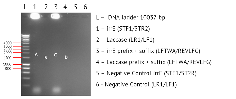
Results: The image below shows a 1% Agarose Gel of an Analytical Restriction Enzyme Digest for Expt 9.2, with a 1000bp ladder. Again we have not obtained any bands. In Lanes 1 and 3 we expected a product corresponding to the size of the irrE gene (1974bp) as indicated by A and C respectively. C would be marginally larger because it also contains the standard BioBrick prefix and suffix, but this different would not be noticeable on a gel. In Lane 2 and 4 we would expect products corresponding to the size of the Laccase gene (1554bp), as shown by B and D. Again we would expect D to be a slightly larger product because the primers were designed to include the prefix and suffix, but this is not a difference we would expect to detect. The strong patches of white demonstrate to us that our DNA template is at a high concentration, and should be diluted before we attempt any repeats. The lack of any products suggest we need to reconsider the protocol. Revising the annealing temperature or designing new primers will have to be considered.

Conclusion: We will revise the protocol to see if we can detect any bands.
Wednesday 15.08.12
Aim - Repeat the PCR again to optimise results: As an improvement we are using: - Phusion polymerase - Diluting the IrrE DNA 10-fold, due to the excessive dose on DNA shown on the gel from yesterday. - Changing the annealing temperature from 55C to 57C
Method
Step 1 - Setting up PCR tubes: Thaw reagents and add to PCR tubes in the proportions described in the table below
| PCR Components | Volume (ul) |
|---|---|
| 5x Reaction Buffer | 10 |
| 25mM MgCl2 | 4 |
| 10mM dNTPs | 1 |
| 10uM Forward primer | 5 |
| 10uM Reverse primer | 5 |
| DNA Polymerase | 0.25 |
| Nuclease Free Water | 24.25 |
| Template DNA | 0.5 |
| Total Volume | 0.5 |
Step 2 - PCR program: Add PCR tubes to a thermocycler and run under the following conditions.
| PCR conditions | Temp (oC) | Time (s) |
|---|---|---|
| Initial Denaturation (1 cycle) | 95 | 30 |
| Denaturation/Annealing/Extension (30 cycles) | 95/55/72 | 10/25/120 |
| Final Extension (1 cycle) | 72 | 600 |
| Hold | 4 | ∞ |
Step 1 - Setting up PCR tubes: The polymerase used was Phusion, and we had two template DNAs – irrE and Laccase.
| DNA Template | Function | Module | Primer Pair | Primer | Primer Sequence |
|---|---|---|---|---|---|
| BBa_K729002 | Laccase Gene | Degradation | LR1/LF1 | LR1 | gaatacggtctttttataccg |
| LF1 | gaaataactatgcaacgtcg | ||||
| REVLF2/LFTW0 | REVLF2 | gtttcttcctgcagcggccgctactagtagaatacggtctttttataccg | |||
| LFTW0 | gtttcttcgaattcgcggccgcttctagaggaaataactatgcaacgtcg | ||||
| No Template (Negative Control) | N/A | Degradation | LR1/LF1 | LR1 | gaatacggtctttttataccg |
| LF1 | gaaataactatgcaacgtcg | ||||
| BBa_K729001 | irrE | Salt Tolerance | STF1/ST2R | STF1 | atggggccaaaagctaaagctgaagcc |
| ST2R | tcactgtgcagcgtcctgcg | ||||
| STF3/ST4R | STF3 | gtttcttcgaattcgcggccgcttctagagatggggccaaaagctaaagctgaagcc | |||
| ST4R | gtttcttcctgcagcggccgctactagtatcactgtgcagcgtcctgcg | ||||
| No Template (Negative Control) | N/A | Salt Tolerance | STF1/STF2 | STF1 | atggggccaaaagctaaagctgaagcc |
| ST2R | tcactgtgcagcgtcctgcg |
Step 2 – PCR Program: An annealing temperature of 57oC was used instead of the standard 55oC.
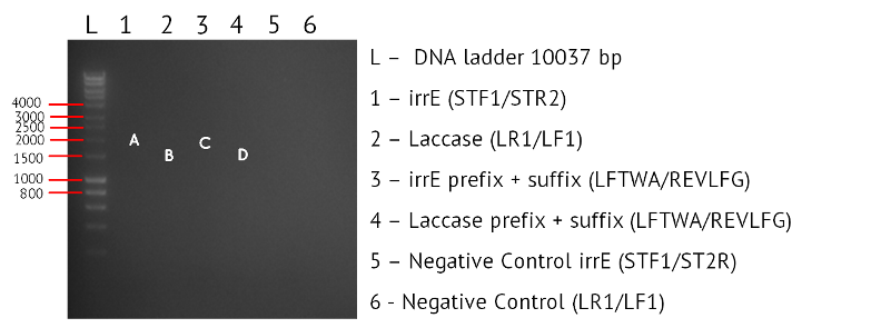
Results: The image below shows a 1% Agarose Gel of an Analytical Restriction Enzyme Digest for Expt 9.2, with a 1000bp ladder. Again we have not obtained any bands. A and C correspond to the bands expected for Laccase, and B and D for bands expected for IrrE. The final two columns are negative controls for which no product was expected. The reason for the failure of this gel is unclear.


Monday 13.8.12
Aim - Check the results of the weekend culture of plates: At the end of last week we set up Expt 9.4, to assess whether poor activity of our antibiotics was a cause of the poor results we get after colony picking. We proposed that our antibiotics may be weak, allowing opportunistic colonies to grow on agar. Therefore we set up a series of agar plates with various antibiotics and controls, to see if we could prove that bacteria without resistance is capable of growing on antibiotic inoculated agar. If this is the case, we know our antibiotic is weak.

Monday 13.8.12
1. Prepare 11-16 plates (10ml DNAse - Agar + 10ul AMP + 10ul 1M IPTG). IPTG induces the lac promoter which in turn activates the transcription of nuclease.
2. Streak cells onto all plates at the same time
3. Incubate at 37°C
4. Apply hydrochloric acid (HCl) to the first plate before putting in the incubator (set time as zero)
5. Take a second reading after four hours, followed by six readings every 3 hours, and a final three readings every two hours.
6. When the reading is taken, observe the following:
a) Diameter of the colony (once the diameter of the colony is measured, pick the colony and put it to grow in LB for nine hours)
b) Diameter of the halo that is achieved once HCl is applied
c) OD from a)
d) Estimate of the depth of the colony on the agar plate
For BL21 cell line that has been modified to contain nuclease:
1. Prepare 11-16 plates (DNAse - Agar + CMP)
2. Streak cells onto all plates at the same time
3. Incubate at 37°C
4. Apply HCl to the first plate before putting in the incubator (set time as zero)
5. Take a second reading after four hours, followed by six readings every 3 hours, and a final three readings
every two hours.
6. When the reading is taken, observe the following:
a. Diameter of the colony (once the diameter of the colony is measured, pick the colony and put it to grow in LB for nine hours)
b. Diameter of the halo that is achieved once HCL is applied
c. OD from a)
d. Estimate of the depth of the colony on the agar plate
Conclusion - Our WNu cell are producing nuclease detectable by this assay, allowing us to conclude that this protocol is sufficient.

Monday 13.8.12
Aims – To test the growth of marine bacteria in different media: This is to optimise the conditions of growth for the bacteria. A test for the marine bacteria will be compared to control E.coli, and the difference in growth measured using a spectrometer. The two media being tested are LB media (typically used for E.coli) and marine media. We expect to have greater optical density of indoliflex in marine medium & greater optical density for W3110 in LB.
Methods
Step 2 - Inoculating Colonies into a Selective Broth:: Add Yul of antibiotic to reach desired antibiotic concentration.
(For Ampicillin this is 50ug/ml, For Kanamycin it is 25ug/ml, for Tetracycline it is 15ug/ml, and for Chloramphenicol it is 25ug/ml)
Step 4 – Selecting a Colony: Select a clear, isolated colony and using an inoculation hoop scoop up a colony onto the tip. Deposit in the falcon tube
Step 5 - Culture: Culture your falcon tubes overnight at a temperature of 37 oC. Leave for no longer than 16 hours.
Step 2 – Inoculating Colonies into a Selective Broth: The table below indicates the volume of broth and the concentration of antibiotic required for each BioBrick. Each cell type will be inoculated into both types of media.
| Type of Bacterium | LB media | Marine media | ||
|---|---|---|---|---|
| No. Falcons | Volume Used (ml) | No. Falcons | Volume Used | |
| W3110 (control) | 3 | 2 | 1 | 2 |
| Oceanibulbus indoliflex (marine) | 2 | 2 | 4 | 2 |
Tuesday 14.8.12
Aim: To measure the optical density of bacterium after overnight culture in different media.
Method?
Results: The table below summarises the results of OD600 measurement. As expected, all three samples of W3110 grown in LB media have a higher optical density (and therefore growth) than the single sample of W3110 grown in marine media. The marine bacterium Oceanibulbus indoliflex also gave us our expected results, with the four samples grown in marine media giving higher optical density (and therefore growth) than the two samples grown in LB media.
| Type of bacterium | Optical Density 600 in LB media | Optical Density 600 in Marine media | ||||||
|---|---|---|---|---|---|---|---|---|
| Sample 1 | Sample 2 | Sample 3 | Sample 1 | Sample 2 | Sample 3 | Sample 4 | ||
| W3100 (Control | Run 1 | 0.526 | 1.085 | 1.035 | 0.185 | / | / | / |
| Run 2 | 0.525 | 1.082 | 1.023 | 0.184 | / | / | / | |
| Run 3 | 0.526 | 1.078 | 1.021 | 0.183 | / | / | / | |
| Oceanibulbus indoliflex (Marine) | Run 1 | 0.033 | 0.003 | / | 0.291 | 0.239 | 0.335 | 0.325 |
| Run 2 | 0.033 | 0.003 | / | 0.289 | 0.226 | 0.335 | 0.322 | |
| Run 3 | 0.033 | 0.003 | / | 0.290 | 0.226 | 0.336 | 0.320 | |
Conclusions
This demonstrates the better suitability of marine media for the marine bacterium. Glycerol stocks were made of the W3110 Sample 2 cultured in LB media, and of the Oceanibulbus indoliflex Sample 3 culture in marine media, as these had the highest density. The W3110 sample will be used for competent cell preparation.
 "
"