Team:University College London/LabBook/Week8
From 2012.igem.org
Sednanalien (Talk | contribs) (→Wednesday 1.8.12) |
Sednanalien (Talk | contribs) (→Tuesday 31.8.12) |
||
| Line 160: | Line 160: | ||
|- | |- | ||
|} | |} | ||
| + | |||
== Tuesday 31.8.12 == | == Tuesday 31.8.12 == | ||
| Line 207: | Line 208: | ||
| 90ul | | 90ul | ||
|} | |} | ||
| - | |||
| - | |||
== Wednesday 1.8.12 == | == Wednesday 1.8.12 == | ||
Revision as of 01:21, 27 September 2012


Monday 30.6.12
Aim: Repeat the gel from Expt 7.3 (week 7) with a smaller ladder and higher agarose percentage in order to detect the very small inserts of BBa_J23119 (35bp) and BBa_B0034 (12hp). Previous ladder used was 10037 bp, and this time we used a 25bp ladder.
Step 1 - Thawing cells: Thaw all materials on ice
Step 2 - Adding Ingredient: Add the following ingredients to autoclaved/sterile eppendorf tubes
| Component | Amount (ul) (one enzyme used) | Amount (ul) (two enzymes used) |
|---|---|---|
| dH20 | 2.5 | 1.5 |
| Buffer 1x | 1 | 1 |
| DNA template | 5 | 5 |
| BSA | 0.5 | 0.5 |
| Enzyme 1 | 1 | 2 |
| Enzyme 2 | N/A | 1 |
Step 3 - Addition of BioBrick: Flick contents gently and centrifuge.
Step 4 - Centrifuge:
RPM: 14000
Time: 1 minute
Temperature: 18oC
Step 5 - Digest Program: Place the samples on a thermocycler under the following conditions:
RPM: 550
Time: 2.5 hours
Temperature: 37oC
Step 6 - Denaturing Enzymes: If you are not running the samples on a gel immediately, denature the restriction enzymes by running the samples on a thermocycler under the following conditions:
RPM: 550
Time: 25 minutes
Temperature: 65oC
Step 2 - Setting up Digests and Controls: The protocol describes the recipe for (i) Digested Plasmid and (ii) Uncut Control. The table below indicates that an uncut and an Xba1/Spe1 digested sample be set up for each BioBrick. Set up Eppendorfs as follows
| Samples | Recipe | Enzymes | |
|---|---|---|---|
| BioBrick | BBa_J23119 | Digested Plasmid | Xba1 & Spe1 |
| Undigested Plasmid (Control) | None | ||
| BBa_B0034 | Digested Plasmid | Xba1 & Spe1 | |
| Undigested Plasmid (Control) | None | ||
Preparing the Gel
Step 1: Within a conical flask, add 3ml 50X TAE, 1.5g Agarose, and 150ml RO water
Step 2: Heat for 1 min in microwave. Swirl. Heat again for 30s. If solution is clear stop. Else repeat.
Step 3: Cool solution under running cold water.
Step 4: Add 20ul ethidium bromide (normal concentration of EB solution is 500ug/ul)
Step 5: Pour into a sealed casting tray in a slow steady stream, ensuring there are no bubbles
Running a gel
Step 6: Add 1 part loading buffer to five parts of loading sample
Step 7: Position the gel in the tank and add TAE buffer, enough to cover the gel by several mm
Step 8: Add 5ul of DNA ladder to lane 1
Step 9: Add samples to the remaining wells
Step 10: Run at 100 volts for 1hour and 15 minutes
Imaging the Gel
Step 11: Place gel in GelDoc 2000 chamber
Step 12: Turn GelDoc 2000 chamber on
Step 13: From computer: Quantity One > Scanner > Gel_Doc_Xr>Manuqal Acquire
Step 14: Alter the exposure/settings to give a clear image.
TAE - Tris-acetate-EDTA
EDTA - ethylenediamine tetraacetic acid
Results:The image below shows a 3.5% Agarose Gel of an Analytical Restriction Enzyme Digest for Expt 7.3. The high agarose concentration of the gel was intended to slow the progression of DNA fragments to enable us to detect the small inserts of BBa_J23119 and BBa_B0034. The table below indicates the expected sizes of BioBricks and Plasmid Backbones. Accordingly, we used a the smallest available ladder, with fragments ranging from 25bp to 500bp. At the top of the gel it is possible to see the large fragments we detected on a 1000bp gel in Expt 7.3 Week 7. However, we had expected this gel to also demonstrate the BBa_J23119 insert (35bp) corresponding to letter A in Lane 1, and the BBa_B0034 insert, corresponding to letter B in Lane 3. No product was found. As expected, there was no product measurable against the 25bp ladder for the uncut plasmids in Lane 2 and Lane 4.

| BioBrick | Expected BioBrick Size (bp) | Plasmid Backbone | Plasmid Expected Size (bp) | Combined Size |
|---|---|---|---|---|
| BBa_J23119 | 35 | PSB1A2 | 2079 | 2114 |
| BBa_B0034 | 12 | PSB1A2 | 2079 | 2091 |
Conclusion:No product was detected for either BioBrick insert. However, we do no feel this necessarily proves the transformation failed, especially as we detected the correct plasmid backbone for each in week 7 (Expt 7.3). The modest nanodrop concentration obtained last week suggests that the concentration was not high enough to allow such a small fragment to be visualised. We are considering alternatives such as running a polyacrylamide gel, which is more sensitive to small fragments, or amplifying the fragment using PCR

Monday 30.7.12
Aim - Picking colonies: Colonies were picked for BBa_I750016, which demonstrated colony formation in Expt 7.4 on Friday 27.7.12.
Step 2 - Inoculating Colonies into a Selective Broth:: Add Yul of antibiotic to reach desired antibiotic concentration.
(For Ampicillin this is 50ug/ml, For Kanamycin it is 25ug/ml, for Tetracycline it is 15ug/ml, and for Chloramphenicol it is 25ug/ml)
Step 4 – Selecting a Colony: Select a clear, isolated colony and using an inoculation hoop scoop up a colony onto the tip. Deposit in the falcon tube
Step 5 - Culture: Culture your falcon tubes overnight at a temperature of 37 oC. Leave for no longer than 16 hours.
Step 2 – Inoculating Colonies into a Selective Broth: The table below indicates the volume of broth and the concentration of antibiotic required for each BioBrick.
| Samples | Volume Inoculated | Broth (ml) | Antibiotic (ug/ml) | |
|---|---|---|---|---|
| BioBrick | BBa_I750016 | 10ul | Lysogeny Broth (5) | Ampicillin(50ug/ml) |
| 90ul | ||||
Tuesday 31.7.12
Aim – Results from Colony Picking
Results: The table below indicates whether there was growth for BBa_I750016
| Samples | Volume Inoculated | Colony Formation | |
|---|---|---|---|
| BioBrick | BBa_I750016 | 10μl | Yes |
| 90μl | Yes | ||
Conclusion: We can proceed onto Miniprep, Analytical Digest, and Nanodrop.
Method
Miniprep:
Step 1 - Pellet Cells: Pellet 1.5-5ml bacterial culture containing the plasmid by centrifugation g = 6000
Time = 2 min
Temperature = (15-25oC)
Step 2 - Resuspend Cells: Add 250ul S1 to the pellet and resuspend the cells completely by vortexing or pipetting. Transfer the suspension to a clean 1.5ml microcentrifuge tube.
Step 3 - Puncturing Cell Membrane: Add 250ul S2 gently mix by inverting the tube 4-6 times to obtain a clear lysate. Incubate on ice or at room temperature for NOT longer than 5 min.
Step 4 - Neutralising S2: Add 400ul Buffer NB and gently mix by inverting the tube 6-10 times, until a white precipitate forms.
Step 5 - Centrifuge:
g: 14000
Time:10 minutes
Temperature: (15-25oC)
Step 6 - Centrifuge: Transfer the supernatant into a column assembled in a clean collection tube (provided. Centrifuge:
g = 10,000
Time: 1 minute
Temperature: (15-25oC)
Step 7 - Wash Column: Discard the flow-through and wash the spin column by adding 700ul of Wash Buffer. Centrifuge:
g - 10,000
Time - 1 minute
Temperature: (15-25oC)
Step 8 - Remove residual ethanol: Centrifuge:
g - 10,000
Time - 1 minute
Temperature: (15-25oC)
Step 9 - Elute DNA: Place the column into a clean microcentrifuge tube. Add 50-100ul of Elution Buffer, TE buffer or sterile water directly onto column membrane and stand for 1 min. Centrifuge:
g - 10,000
Time - 1 minute
Temperature: (15-25oC)
Step 10 - Storage: Store DNA at 4oC or -20oC
Wednesday 1.8.12
Aim: Nanodrop of BBa_I750016
| BioBrick | λ260 | λ 280 |
|---|---|---|
| BBa_I750016 (ng/μl) | 7.6 | 7.6 |
Conclusion: The concentration of BioBrick is to low to justify running a gel. We conclude the transformation failed.

Monday 30.7.12
Aim - Transformation of previously failed BioBricks: BBa_C0040 was transformed in Expt 7.4, but appeared to be contaminated. BBa_R0040, as part of the same experiment, did not form colonies at all after transformation. Therefore both were reattempted.
Method:
Step 1 - Addition of BioBrick: To the still frozen competent cells, add 1 - 5 µL of the resuspended DNA to the 2ml tube.
Step 4 - Incubation: Close tube and incubate the cells on ice for 45 minutes.
Step 5 - Heat Shock: Heat shock the cells by immersion in a pre-heated water bath at 37ºC for 10 minutes.
Step 6 - Incubation: Incubate the cells on ice for 2 minutes.
Step 7 - Add media: Add 1.5ml of Lysogeny Broth and transfer to a falcon tube.
Step 8 - Incubation: Incubate the cells at 37ºC for 1 hour at RPM 550.
Step 9 - Transfer: transfer the solution back into a 1.5ml Eppendorf and centrifuge
RPM: 14000
Time: 2 minutes
Temperature (18-25oC)
Step 10 - Resuspend:Remove supernatant and resuspend in 100ul LB
Step 11 - Plating: Spread the resuspended cell solution onto a selective nutrient agar plate. Place the plates in a 37°C static incubator, leave overnight (alternatively a 30°C static incubator over the weekend)
Step 1 – Thawing Cells: Use W3110 cell line created in Week 2 (Expt 2.1)
Step 3 – Addition of BioBrick: To each 2ml eppendorf, add 1ul of the following BioBricks. Include an extra tube as a control, with no BioBrick added
| Function | Module | ||
|---|---|---|---|
| BioBrick | BBa_C0040 | Tetracycline Repressor | Buoyancy |
| BBa_R0040 | TetR Repressible Promoter | Buoyancy | |
| Control | Positive (one for each of the above BioBricks) | ||
| Negative (No BioBrick) | |||
Step 9 – Plating samples on Agar Plates: The table below indicates the chosen inoculation volume (two for each BioBrick) and the correct gel antibiotic concentration for all samples.(Extra caution was taken to allow agar to cool before adding Ampicillin, in case this is the cause of difficulty).
| Samples | Volume Inoculated | Antibiotic in Gel (ug/ml) | |
|---|---|---|---|
| BioBrick | BBa_C0040 | 10ul | Ampicillin(50ug/ml) |
| 90ul | |||
| BBa_R0040 | 10ul | ||
| 90ul | |||
| Control | Positive (Contains BioBrick BBa_C0040) | 36ul | No Antibiotic |
| Negative (No BioBrick) | 36ul | 1x Ampicillin(50ug/ml) | |
Tuesday 31.8.12
Aim - Check results of Transformation: The table below indicates whether there was growth on the Agar Plates after Transformation.
| Samples | Volume Inoculated | Colony Formation | |
|---|---|---|---|
| BioBrick | BBa_C0040 | 10ul | Yes |
| 90ul | Yes | ||
| BioBrick | BBa_R0040 | 10ul | Yes |
| 90ul | Yes | ||
| Control | Positive (Contains BioBrick BBa_C0040) | 36ul | Yes |
| Negative (No BioBrick) | 36ul | No | |
Conclusion: Transformation was successful so will proceed to Colony Picking
Method
Aim - Picking colonies:
Step 2 - Inoculating Colonies into a Selective Broth:: Add Yul of antibiotic to reach desired antibiotic concentration.
(For Ampicillin this is 50ug/ml, For Kanamycin it is 25ug/ml, for Tetracycline it is 15ug/ml, and for Chloramphenicol it is 25ug/ml)
Step 4 – Selecting a Colony: Select a clear, isolated colony and using an inoculation hoop scoop up a colony onto the tip. Deposit in the falcon tube
Step 5 - Culture: Culture your falcon tubes overnight at a temperature of 37 oC. Leave for no longer than 16 hours.
Step 2 – Inoculating Colonies into a Selective Broth: The table below indicates the volume of broth and the concentration of antibiotic required for each BioBrick.
| Samples | Volume Inoculated | Broth (ml) | Antibiotic (ug/ml) | |
|---|---|---|---|---|
| BioBrick | BBa_C0040 | 10ul | Lysogeny Broth (5) | Ampicillin(50ug/ml) |
| 90ul | ||||
| BBa_R0040 | 10ul | |||
| 90ul | ||||
Wednesday 1.8.12
Aim – Results from Colony Picking
Results: The table below indicates whether there was growth for the BioBricks
| Samples | Volume Inoculated | Growth | |
|---|---|---|---|
| BioBrick | BBa_C0040 | 10μl | Yes |
| 90μl | Yes | ||
| BBa_R0040 | 10μl | Yes | |
| 90μl | Yes | ||
Conclusion: We can move on to miniprep
Methods
Miniprep of Samples
Step 1 - Pellet Cells: Pellet 1.5-5ml bacterial culture containing the plasmid by centrifugation g = 6000
Time = 2 min
Temperature = (15-25oC)
Step 2 - Resuspend Cells: Add 250ul S1 to the pellet and resuspend the cells completely by vortexing or pipetting. Transfer the suspension to a clean 1.5ml microcentrifuge tube.
Step 3 - Puncturing Cell Membrane: Add 250ul S2 gently mix by inverting the tube 4-6 times to obtain a clear lysate. Incubate on ice or at room temperature for NOT longer than 5 min.
Step 4 - Neutralising S2: Add 400ul Buffer NB and gently mix by inverting the tube 6-10 times, until a white precipitate forms.
Step 5 - Centrifuge:
g: 14000
Time:10 minutes
Temperature: (15-25oC)
Step 6 - Centrifuge: Transfer the supernatant into a column assembled in a clean collection tube (provided. Centrifuge:
g = 10,000
Time: 1 minute
Temperature: (15-25oC)
Step 7 - Wash Column: Discard the flow-through and wash the spin column by adding 700ul of Wash Buffer. Centrifuge:
g - 10,000
Time - 1 minute
Temperature: (15-25oC)
Step 8 - Remove residual ethanol: Centrifuge:
g - 10,000
Time - 1 minute
Temperature: (15-25oC)
Step 9 - Elute DNA: Place the column into a clean microcentrifuge tube. Add 50-100ul of Elution Buffer, TE buffer or sterile water directly onto column membrane and stand for 1 min. Centrifuge:
g - 10,000
Time - 1 minute
Temperature: (15-25oC)
Step 10 - Storage: Store DNA at 4oC or -20oC
Nanodrop
Software ND-1000 Model:
Step 1: Initialise the spectrophotometer by pipetting 1 µ of clean water onto lower optic surface, lowering the lever arm and selecting ‘initialise’ in the ND-1000 software
Step 2: Wipe and add elution buffer as negative control. Click blank in ND-1000 software
Step 3: Wipe and add 1 µl sample
Step 4: On the software set lambda to 260nm
Step 5: Lower the lever arm and click measure in ND-1000 software
Step 6: Take readings for concentration and purity
Step 7: Once measurement complete, wipe surface
Results:
| BioBrick | λ260 | λ 280 |
|---|---|---|
| BBa_C0040 (ng/μl) | 9.4 | 18.9 |
| BBa_R0040 (ng/μl) | 9.3 | 36.9 |
Conclusion: The concentration of these plasmids is very low. We can conclude that the transformation has failed. The reasons for the continued failure of these plasmids are unclear

Tuesday 31.7.12
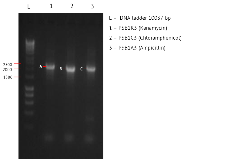
Aim: to generate enough plasmid backbones (pSB1K3, pSB1C3, pSB1A3) that we could use for 3A ligation. After the PCR reaction, we can detect the presence of the correct products by running a 1% gel electrophoresis.
Method:
Step 1 - Setting up PCR tubes: Thaw reagents and add to PCR tubes in the proportions described in the table below
| PCR Components | Volume (ul) |
|---|---|
| 5x Reaction Buffer | 10 |
| 25mM MgCl2 | 4 |
| 10mM dNTPs | 1 |
| 10uM Forward primer | 5 |
| 10uM Reverse primer | 5 |
| DNA Polymerase | 0.25 |
| Nuclease Free Water | 24.25 |
| Template DNA | 0.5 |
| Total Volume | 0.5 |
Step 2 - PCR program: Add PCR tubes to a thermocycler and run under the following conditions.
| PCR conditions | Temp (oC) | Time (s) |
|---|---|---|
| Initial Denaturation (1 cycle) | 95 | 30 |
| Denaturation/Annealing/Extension (30 cycles) | 95/55/72 | 10/25/120 |
| Final Extension (1 cycle) | 72 | 600 |
| Hold | 4 | ∞ |
Wednesday 1.8.12
Aim: to run a 1% gel of the products of yesterdays PCR reaction.
Method
Preparing the Gel
Step 1: Within a conical flask, add 3ml 50X TAE, 1.5g Agarose, and 150ml RO water
Step 2: Heat for 1 min in microwave. Swirl. Heat again for 30s. If solution is clear stop. Else repeat.
Step 3: Cool solution under running cold water.
Step 4: Add 20ul ethidium bromide (normal concentration of EB solution is 500ug/ul)
Step 5: Pour into a sealed casting tray in a slow steady stream, ensuring there are no bubbles
Running a gel
Step 6: Add 1 part loading buffer to five parts of loading sample
Step 7: Position the gel in the tank and add TAE buffer, enough to cover the gel by several mm
Step 8: Add 5ul of DNA ladder to lane 1
Step 9: Add samples to the remaining wells
Step 10: Run at 100 volts for 1hour and 15 minutes
Imaging the Gel
Step 11: Place gel in GelDoc 2000 chamber
Step 12: Turn GelDoc 2000 chamber on
Step 13: From computer: Quantity One > Scanner > Gel_Doc_Xr>Manuqal Acquire
Step 14: Alter the exposure/settings to give a clear image.
TAE - Tris-acetate-EDTA
EDTA - ethylenediamine tetraacetic acid
Results: The image below shows a 1% Agarose Gel of an Analytical Restriction Enzyme Digest for Expt 8.2. Lane 1, 2 and 3 all show products of approximately 2000bp which is of the expected size for the plasmid backbones PSB1K3 (indicated by A), PSB1C3 (indicated by B) and PSB1A3 (indicated by C).

| Plasmid Backbone | Expected Size bp |
|---|---|
| PSB1K3 (Kanamycin) | 2000 |
| PSB1C3 (Chloramphenicol) | 2000 |
| PSB1A3 (Ampicillin) | 2000 |
Conclusion:The PCR was successful.
Friday 3.8.12
Aim: PCR clean up for the plasmid backbones and nanodrop.
Methods:
PCR clean up
Software ND-1000 Model:
Step 1: Initialise the spectrophotometer by pipetting 1 µ of clean water onto lower optic surface, lowering the lever arm and selecting ‘initialise’ in the ND-1000 software
Step 2: Wipe and add elution buffer as negative control. Click blank in ND-1000 software
Step 3: Wipe and add 1 µl sample
Step 4: On the software set lambda to 260nm
Step 5: Lower the lever arm and click measure in ND-1000 software
Step 6: Take readings for concentration and purity
Step 7: Once measurement complete, wipe surface
Results: Nanodrop concentrations are presented in the table below. All are low except for the backbone containing kanamycin. We concluded that PCR for these backbones needs to be repeated
| BioBrick | λ260 | λ 280 |
|---|---|---|
| PSB1A3 (ng/μl) | 27.3 | 26.6 |
| PSB1C3 (ng/μl) | 23 | 24.4 |
| PSB1K3 (ng/μl) | 66.5 | 87 |
Conclusion: The PCR for PSB1A3 and PSB1C3 need to be repeated.

Tuesday 31.7.12
Aim: In order to set up our salt tolerance characterisation we must grow up 10ml of W3110 and Salt Tolerance (BBa_K398108) transformed W3110 overnight
Wednesday 1.8.12
Aim: Today we commence the protocols for characterising salt tolerance.
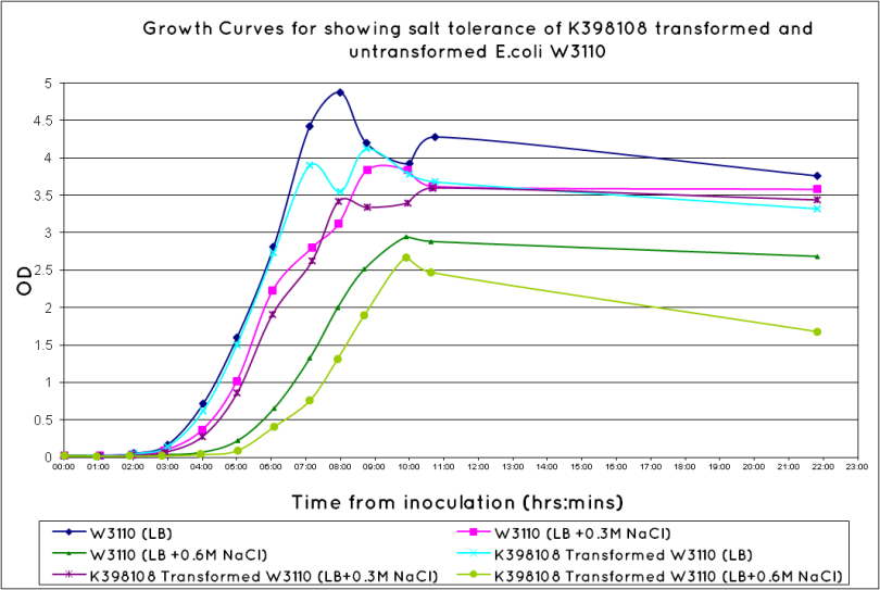
Results: Our results demonstrate that out transformed cells are salt tolerant, compared to the untransformed W3110 cells.Our results reflect those of TU Delft 2010 who characterised the same BioBrick. However they did not show their results beyond the exponential phase, which we have included.


Conclusions:
Growth rate during exponential phase is greater in the K398108 transformed E.coli W3110 than in the untransformed (same conclusion as drawn by TU Delft 2010). Increase in growth rates significantly lower than original at highest salt concentration – could be explained by use of different E.coli strains i.e. higher growth rate of the non-transformed strain. We plan to repeat to strengthen conclusions.

Wednesday 1.8.12
Aim: Start 3A assembly to ligate the first (BBa_J23119+BBa_B0034), and second (plasmid backbone PSB1K3 and Pcst starvation promoter) constructs.
Method
Thursday 2.8.12
Aim: Evaluation of the plates from 3A ligation. This should inform us as to whether we can proceed and do the colony picking of both A (J23119+BB0034) and B (Pcst + BB0034) constructs.
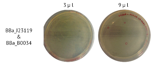
Result: Observations showed that 3A was successful for the construct A, as shown in the image below. However there was no growth assembled pCST+BBa_B0034 (construct B). The following table summarises the results.

Conclusion: Colony picking was carried out for the first construct (BBa_J23119+BBa_B0034), but not from the second construct (plasmid backbone PSB1K3 and Pcst starvation promoter).
Method
Step 2 - Inoculating Colonies into a Selective Broth:: Add Yul of antibiotic to reach desired antibiotic concentration.
(For Ampicillin this is 50ug/ml, For Kanamycin it is 25ug/ml, for Tetracycline it is 15ug/ml, and for Chloramphenicol it is 25ug/ml)
Step 4 – Selecting a Colony: Select a clear, isolated colony and using an inoculation hoop scoop up a colony onto the tip. Deposit in the falcon tube
Step 5 - Culture: Culture your falcon tubes overnight at a temperature of 37 oC. Leave for no longer than 16 hours.
Friday 3.8.12
Aim:Check the results of colony picking.
Results: There was growth. Next we can proceed to run a gel and a nanodrop to detect the presence of the correct bands, and whether we have a useable concentration.

Thursday 2.8.12
Aim - Transformation of alternative BioBricks: We have colony picked the BBa_J23119 constitutive promoter and the BBa_B0034 Ribosome Binding Site twice (Expt 6.3 & Expt 7,3), and gained a reasonable plasmid concentration. However the concentration was not high enough to be able to detect the product on the gel. This concerns us, because although we have detected the correct plasmid backbone, we are worried that we lack evidence that the transformation was successful. One option is to attempt several other BioBricks with the same function, in order to increase the chances of generating a product on the gel - and confirming that we definitely have a BioBricks with the required function.It would also increase the chance of generating successful ligations if we can reach a higher concentration of plasmid. We will also be reattempting the transformation of BBa_I750016 which failed several attempts at colony picking.
Method:
Step 1 - Addition of BioBrick: To the still frozen competent cells, add 1 - 5 µL of the resuspended DNA to the 2ml tube.
Step 4 - Incubation: Close tube and incubate the cells on ice for 45 minutes.
Step 5 - Heat Shock: Heat shock the cells by immersion in a pre-heated water bath at 37ºC for 10 minutes.
Step 6 - Incubation: Incubate the cells on ice for 2 minutes.
Step 7 - Add media: Add 1.5ml of Lysogeny Broth and transfer to a falcon tube.
Step 8 - Incubation: Incubate the cells at 37ºC for 1 hour at RPM 550.
Step 9 - Transfer: transfer the solution back into a 1.5ml Eppendorf and centrifuge
RPM: 14000
Time: 2 minutes
Temperature (18-25oC)
Step 10 - Resuspend:Remove supernatant and resuspend in 100ul LB
Step 11 - Plating: Spread the resuspended cell solution onto a selective nutrient agar plate. Place the plates in a 37°C static incubator, leave overnight (alternatively a 30°C static incubator over the weekend)
Step 1 – Thawing Cells: Use W3110 cell line created in Week 2 (Expt 2.1)
Step 3 – Addition of BioBrick: To each 2ml eppendorf, add 1ul of the following BioBricks. Include an extra tube as a control, with no BioBrick added
| Function | Module | ||
|---|---|---|---|
| BioBrick | BBa_J23100 | Constitutive Promoter | All |
| BBa_J23106 | Constitutive Promoter | All | |
| BBa_B0030 | Ribosome Binding Site | All | |
| BBa_I750016 | Ribosome Binding Site | All | |
| Control | Positive (one for each of the above BioBricks) | ||
| Negative (No BioBrick) | |||
Step 9 – Plating samples on Agar Plates: The table below indicates the chosen inoculation volume (two for each BioBrick) and the correct gel antibiotic concentration for all samples.(Extra caution was taken to allow agar to cool before adding Ampicillin, in case this is the cause of difficulty).
| Samples | Volume Inoculated | Antibiotic in Gel (ug/ml) | |
|---|---|---|---|
| BioBrick | BBa_J23100 | 10ul | Ampicillin(50ug/ml) |
| 90ul | |||
| BBa_J23106 | 10ul | ||
| 90ul | |||
| BBa_B0030 | 10ul | ||
| 90ul | |||
| BBa_I750016 | 10ul | ||
| 90ul | |||
| Control | Positive | 36ul | No Antibiotic |
| Negative (No BioBrick) | 36ul | 1x Ampicillin(50ug/ml) | |
Friday 3.8.12
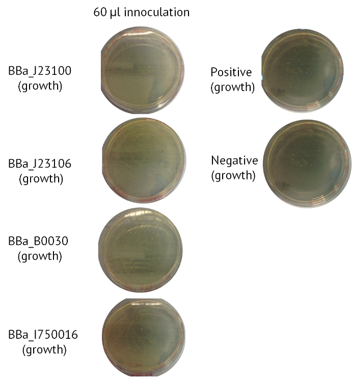
Aim - Check results of Transformation: The table below indicates whether there was growth on the Agar Plates after Transformation. Included below are images of the Agar Plates for each BioBrick.
| Samples | Volume Inoculated | Colony Formation | |
|---|---|---|---|
| BioBrick | BBa_J23100 | 10ul | Yes |
| 90ul | Yes | ||
| BBa_J23106 | 10ul | Yes | |
| 90ul | Yes | ||
| BBa_B0030 | 10ul | Yes | |
| 90ul | Yes | ||
| BBa_I750016 | 10ul | Yes | |
| 90ul | Yes | ||
| Control | Positive (Contains BioBrick BBa_C0040) | 36ul | Yes |
| Negative (No BioBrick) | 36ul | Yes | |

Conclusion: We had growth for all of our BioBricks, but also for the negative control which indicates there was contamination. Given we have had several issues with negative control recently, we decided we would not let the contamination delay the project. We will risk colony picking with the hope that we have some correctly transformed colonies. This will be commenced next week. It is worth noting that for Constitutive Promoter BBa_J23106 and for the Gas Vesicle Cluster BBa_I750016 there was a continuous film of growth. This suggests to us that the agar was not selective enough.
 "
"