Team:UNAM Genomics Mexico/Results/Nanotubes
From 2012.igem.org
| Line 71: | Line 71: | ||
<table border="0" height="150" cellspacing="15" bgcolor="transparent" id="tablecontentbg" cellpadding="10"> | <table border="0" height="150" cellspacing="15" bgcolor="transparent" id="tablecontentbg" cellpadding="10"> | ||
<tr> | <tr> | ||
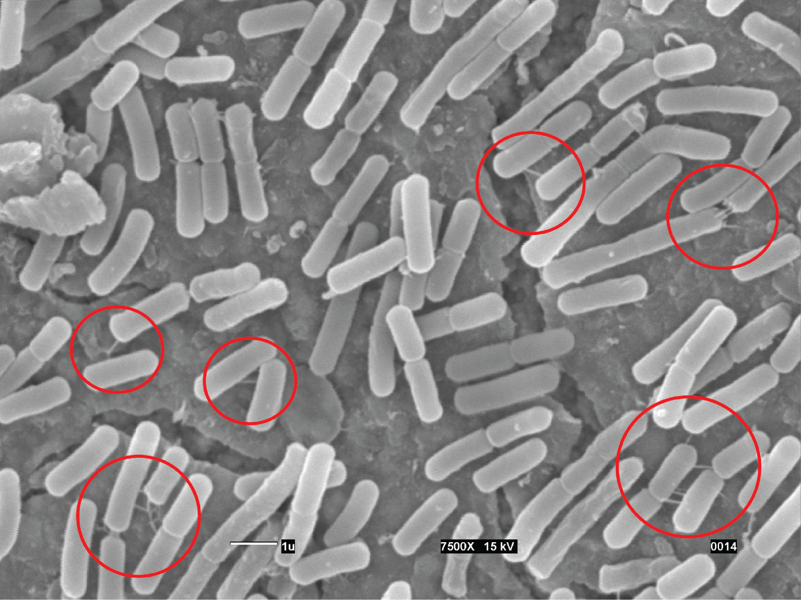
| - | <td id="rightcolumn2" align= "center"><br />[[File:UnamgenomicsNanocirculorojo.jpg|800px| link=Team:UNAM_Genomics_Mexico/Notebook/Bacillus]]<p>'''PY79 cells were grown to midexponential | + | <td id="rightcolumn2" align= "center"><br />[[File:UnamgenomicsNanocirculorojo.jpg|800px| link=Team:UNAM_Genomics_Mexico/Notebook/Bacillus]]<p>'''PY79 cells were grown to midexponential phase, plated on LB agar, incubated for 6 hr at 37°C, and visualized by SEM Rej34 circles: (A) Bacillus subtilis cells(x 7,500). The red circles indicate intercellular nanotubes connecting neighboring cells. The scale bar represents 1 micrometer. '''</p></td> |
| - | phase, plated on LB agar, incubated for 6 hr at 37°C, and visualized by SEM Rej34 circles: (A) Bacillus subtilis cells(x 7,500). The red circles indicate intercellular nanotubes connecting neighboring cells. The | + | |
| - | scale bar represents 1 micrometer. '''</p></td> | + | |
</tr> | </tr> | ||
</table> | </table> | ||
| Line 86: | Line 84: | ||
<table border="0" height="150" cellspacing="15" bgcolor="transparent" id="tablecontentbg" cellpadding="10"> | <table border="0" height="150" cellspacing="15" bgcolor="transparent" id="tablecontentbg" cellpadding="10"> | ||
<tr> | <tr> | ||
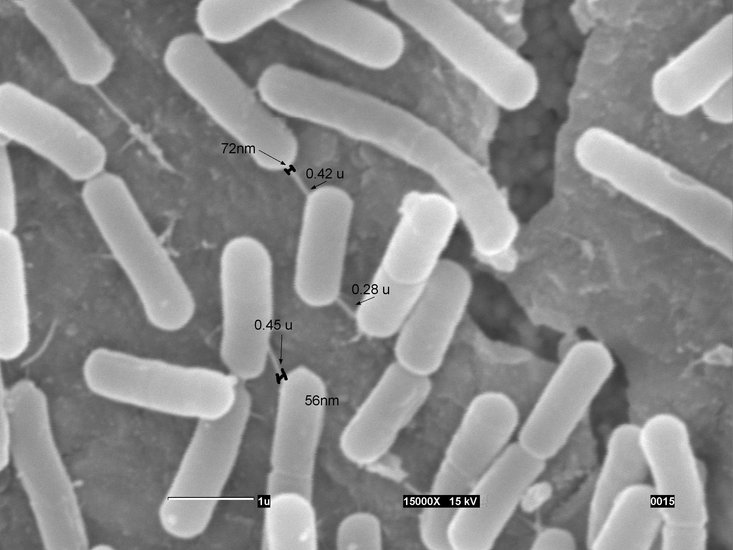
| - | <td id="rightcolumn2" align= "center"><br />[[File:UnamgenomicsREJ35 valores.jpg|800px| link=Team:UNAM_Genomics_Mexico/Notebook/Bacillus]]<p>''' | + | <td id="rightcolumn2" align= "center"><br />[[File:UnamgenomicsREJ35 valores.jpg|800px| link=Team:UNAM_Genomics_Mexico/Notebook/Bacillus]]<p>'''PY79 cells were grown to midexponential phase, plated on LB agar, incubated for 6 hr at 37°C, and visualized by SEM Rej35_valores:(A)Measurment of the length and width of the nanotubes connecting the (PY79) cells grown on solid LB medium. The scale bar represent 1 micrometer'''</p></td> |
</tr> | </tr> | ||
</table> | </table> | ||
| Line 98: | Line 96: | ||
<table border="0" height="150" cellspacing="15" bgcolor="transparent" id="tablecontentbg" cellpadding="10"> | <table border="0" height="150" cellspacing="15" bgcolor="transparent" id="tablecontentbg" cellpadding="10"> | ||
<tr> | <tr> | ||
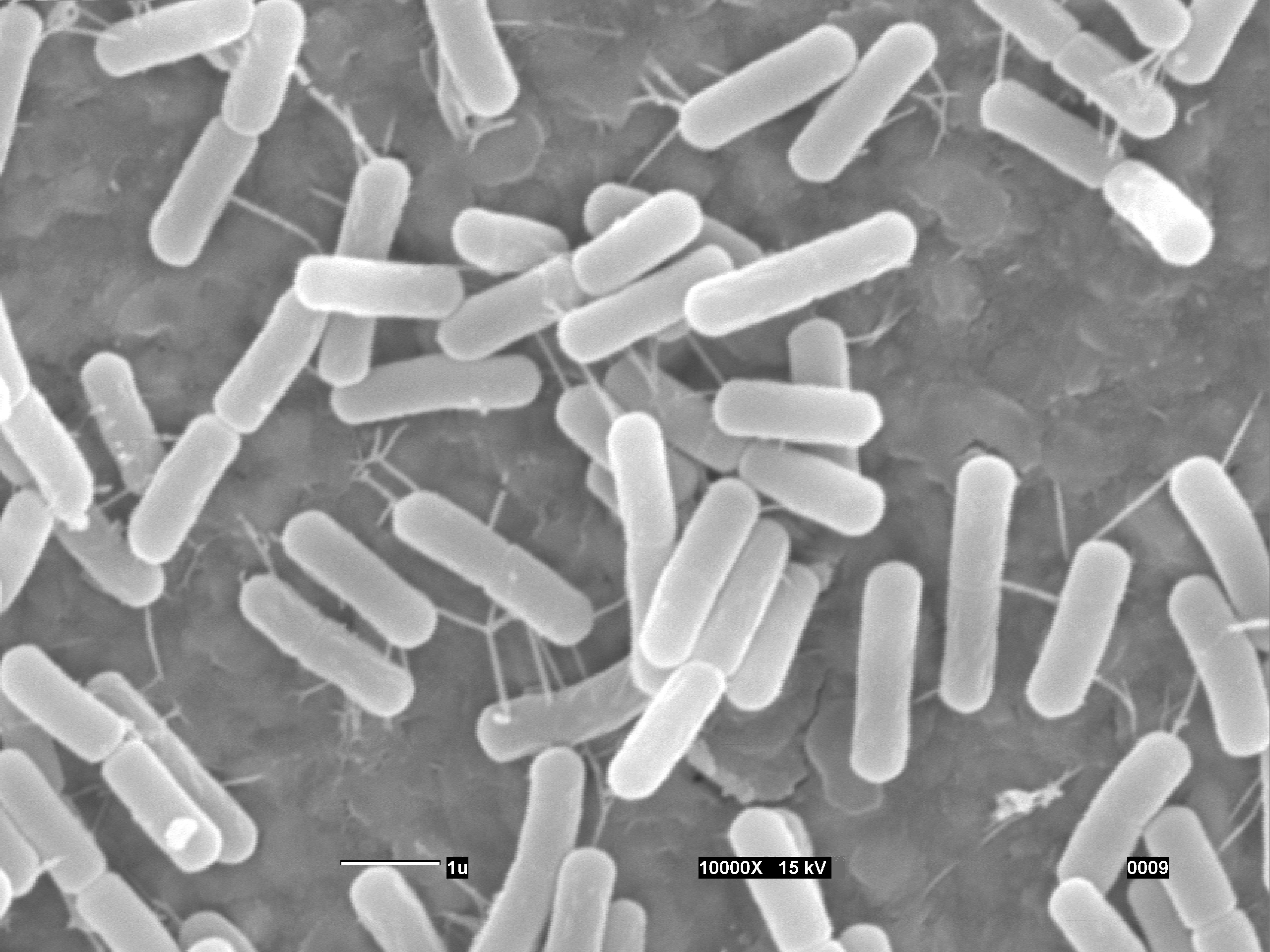
| - | <td id="rightcolumn2" align= "center"><br />[[File:UnamgenomicsREJ48.jpg|800px| link=Team:UNAM_Genomics_Mexico/Notebook/Bacillus]]<p>''' | + | <td id="rightcolumn2" align= "center"><br />[[File:UnamgenomicsREJ48.jpg|800px| link=Team:UNAM_Genomics_Mexico/Notebook/Bacillus]]<p>'''PY79 cells were grown to midexponential phase, plated on LB agar, incubated for 6 hr at 37°C, and visualized by SEM Rej48: (A) Field of cells demonstrating the occurrence of |
| + | a network of intercellular nanotubes (X 10,000). The scale bar represents 1 micrometer'''</p></td> | ||
</tr> | </tr> | ||
</table> | </table> | ||
Revision as of 21:13, 25 October 2012

Nanotubes
 Nanotubes |
 Nanotubes |
 |
Just as people, bacterium need to communicate with others, for this purpose there are many ways, and one of the more recent discovery are Nanotubes that bridge neighboring cells, providing a network for exchange of cellular molecules within and between species. Ben-Yehuda et al discovered these nanotubes in 2011, they show and extraordinary form of communication between Bacillus subtilis. Our team was astonished because of the implications, in the article is described that GFP and calcein, two molecules which cannot leave the cytoplasm, can be transferred to neighboring cells in B. subtillis, it means bacterium can share cytoplasm, the complex network of cells sharing cytoplasm that can be created was our main motivation to create Bacillus booleanus.
It's still little known about nanotubes, and that's make them a very interesting subject of study, and we are not the first iGEM team interested in working with them, in 2011 Paris_Bettencourt team work with the nanotubes, their goal was to characterize them.
To accomplish our goal of communicate our logic gates, we contacted Ben Yehuda group, and Paris Bettencourt iGEM 2011 team, to obtain protocols and work experience.
-

2011 Gyanendra P. Dubey, Sigal Ben-Yehuda. Intercellular Nanotubes Mediate Bacterial Communication. Cell, 2011; 144 (4): 590 DOI:10.1016/j.cell.2011.01.015
-

2011 Gyanendra P. Dubey, Sigal Ben-Yehuda. Intercellular Nanotubes Mediate Bacterial Communication. Cell, 2011; 144 (4): 590 DOI:10.1016/j.cell.2011.01.015
We work to recreate the formation of nanotubes, and we did it!
We prepare our cells with the SEM Analysis Protocol, We fixed our cells in the right moment (6 hrs of growth at 37°C), that is when they are in the exponential growth phase, and then we watch them in the SEM (Scanning Electron Microscope) and we get different and amazing pictures.
We got similar results to the article, tube length ranged from 0.25 micrometers to 0.9 micrometers, whereas width ranged approximately from 50 to 80 nm.
We also got an amazing picture about the nanotubes in which we can see, that there are many nanotubes among the cells,
and that they communicate between different nanotubes to the same cell, observing this amount of nanotubes, we could
think that the probability of transfer will raise.
Please see our wetlab notebook in the clicking the following image:
 OR gate Notebook |
 "
"


