Team:HKUST-Hong Kong/Characterization
From 2012.igem.org
m (Intro Rephrase) |
|||
| Line 366: | Line 366: | ||
<h1>Introduction</h1> | <h1>Introduction</h1> | ||
</div> | </div> | ||
| - | <p>In our project, we have characterized two promoters and the cell death device | + | <p>In our project, we have characterized two promoters and the cell death device using different methods. The results indicate that our parts are functional and we can quantitatively control their activity by changing the experimental conditions.</p> |
</div> | </div> | ||
<div id="paragraph2" class="bodyParagraphs"> | <div id="paragraph2" class="bodyParagraphs"> | ||
Revision as of 13:48, 26 September 2012
Characterization
Introduction
In our project, we have characterized two promoters and the cell death device using different methods. The results indicate that our parts are functional and we can quantitatively control their activity by changing the experimental conditions.
Low Efficiency Constitutive Promoter pTms
Background Information (link to Regulation and Control Module)
Objective
Our objective for characterizing this promoter is to test whether pTms works in E.coli DH10B strain and determine the relative promoter units (RPU) of it to the standard constitutive promoter activity reference point given by part registry so that we and the subsequent IGEM teams may have an idea how efficient this promoter is.
Intended Result
1. pTms should work in E.coli. This is supported by previous research (Moran et al., 1982).
2. The activity of pTms should be relatively low.
Method
Instead of measuring the absolute promoter activity, our characterization was generally based on measuring the relevant in vivo activity of this constitutive promoter. By adopting this method, we may be able to eliminate the error caused by different experimental conditions and give a relatively more convincing result.
By linking the promoter with GFP (BBa_E0240), the promoter activity was represented by the GFP synthesis rate which can be easily measured. E.Coli carrying the right construct was then cultured to log phase. During a time slot around the mid-log phase, the GFP intensity and OD595 value were measured to obtain the Relative Promoter Units (RPU).
Characterization Procedure
- Constructing BBa_K733009-pSB3K3 (pTms-BBa_E0240-pSB3K3); Transforming BBa_I20260-pSB3K3 (Standard Constitutive Promoter Activity Reference Point) from 2012 Distribution;
- Preparing supplemented M9 medium (see below);
- Culturing E.coli DH10B strain carrying BBa_K733009-pSB3K3 and E.coli carrying BBa_I20260-pSB3K3 in supplemented M9 medium and measuring the growth curve respectively;
- Measuring the GFP intensity and ODA595 value every 15 minutes after E.coli carrying BBa_K733009-pSB3K3 and E.coli carrying BBa_I20260 are cultured to mid-log phase;
- Calculating the Relative Promoter Unites (RPU) using the obtained data;
- Compiling the result.
Data Processing
- After E.coli carrying the right construct was grown into mid-log phase, GFP intensity and ODA595 were measured every 15 minutes (up to 60min);
- For GFP intensity, curve reflecting GFP expression change was plotted; for ODA595, average values was taken;
- GFP synthesis rate was then represented by the slope of the curve reflecting GFP expression change;
- Absolute promoter activity of pTms and I20260 were calculated by divide the corresponding GFP synthesis rate by the average ODA595 value;
- Absolute promoter activity was then modified by taking the average value of all sets of data obtained;
- Finally, R.P.U was calculated by dividing pTms absolute promoter activity by I20260 absolute promoter activity.
Result

* GFP expression curve for one set of data
2. The overall RPU was calculated as 0.046497. It has been shown that pTms has a very low promoter efficiency in E.coli DH10B stain.

Discussion
Compared with I20260, it seems that bacteria carrying pTms had a rather low GFP expression. This may cause some difficulty upon deciding whether pTms functions in E.coli or not. However, since viewed from the curve, the GFP expression for pTms increased gradually in respect to time. These all give us good reason to say that pTms functions in E.coli DH10B strain, although with a low efficiency. Another reason for its low efficiency could be that pTms was originally got from B.subtilis and is only suggested to be functional in E.coli. While we haven’t got time to characterize the promoter in B.subtilis, we still hope that in the future we can actually achieve that.
Reference
Moran, C., Lang, N., LeGrice, S., Lee, G., Stephens, M., Sonenshein, A., et al. (1982). Nucleotide sequences that signal the initiation of transcription and translation in Bacillus subtilis..Molecular and General Genetics MGG,186, 339-346.
Supplemented M9 Medium Composition
1. 5X M9 Salt Composition (1L)
(2) 15g KH2PO4
(3) 2.5g NaCl
(4) 5.0g NH4CL
(2) 2ml of 1M MgSO4
(3) 100μl of 1M CaCl2
(4) 5ml of 40% glycerol
(2) 0.2% casamino acids
Xylose Inducible Promoter
Background Information (link to Regulation and Control Module)
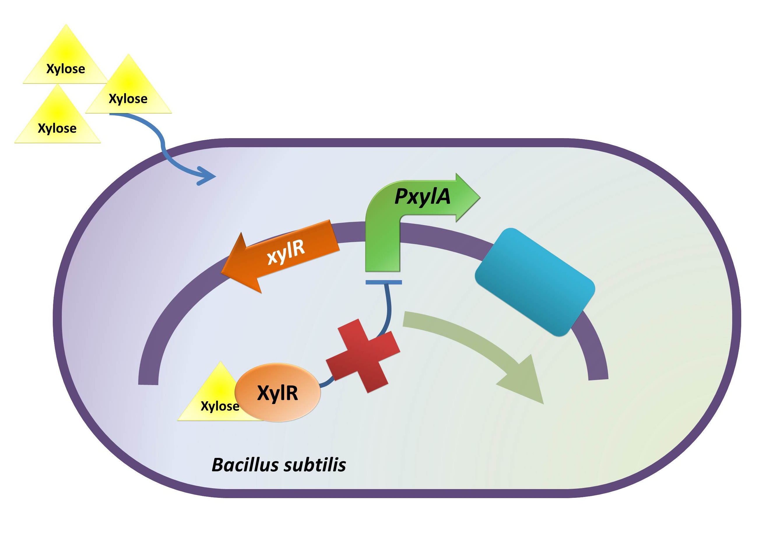
The reason of using xylose inducible promoter is to make the expression of toxin and BMP-2 controllable. Xylose is not toxic and normally is not present in human colon. This provides us an easy way to induce BMP-2 expression without disrupting normal human body function.
Objective
Upon characterizing xylose inducible promoter, we want to test whether xylose inducible promoter works in E.coli DH10B strain and if it works what is the absolute promoter activity under certain experimental condition.
Intended Result
1. Xylose inducible promoter is functional in E.coli.
2. After the inducer concentration has reached certain level, a relatively stationary GFP expression level should be observed.
Method
The absolute promoter activity was measured in respect to induction time and xylose concentration.
Here the same reporter gene (BBa_E0240) was used to indicate promoter activity. E.coli carrying the right construct was cultured to log phase. Following the addition of xylose at serial concentration, during a time slot around the mid log point, the GFP intensity and ODA595 were measured for every 30 min. A curve indicating the GFP intensity unit as a respect of time and xylose concentration was plotted.
Characterization Procedure
- Constructing xylR-PxylA-BBa_E0240-pSB1A2
- Preparing supplemented M9 medium (see below);
- Culturing E.coli carrying xylR-PxylA-BBa_E0240-pSB1A2 and E.coli without constructs in supplemented M9 medium and measuring the growth curve respectively;
- Culturing the same bacteria in supplemented M9 medium to log phase;
- Adding xylose at different concentration to different sets of culture medium;
- Measuring the GFP intensity and OD595 value across time for every sets of culture medium that are of different xylose concentration;
- Plotting a figure about the GFP intensity unit in respect of xylose concentration and time;
- Compiling the result.
Data Processing
- After E.coli carrying the right was growing into mid-log phase, GFP intensity and ODA595 were measured every 30 minutes (up to 120min);
- For GFP intensity, curve reflecting GFP expression change was plotted; for ODA595, average value was taken;
- GFP synthesis rate was then represented by the slope of the curve reflecting GFP expression change;
- Absolute promoter activity for the promoter under different inducer concentrations were calculated by divide the corresponding GFP synthesis rate by the average ODA595 value;
- Absolute promoter activity was then modified by taking the average value of all sets of data obtained.
Result
- Shown by our figure below, under the addition of xylose, GFP expression increased. This tells us that xylose inducible promoter is functional in E.coli DH10B strain.
- When no xylose was added, a little amount of GFP was expressed. This suggests that xylose inducible promoter is to some extent leaky.
- A relatively stationary GFP expression was observed after xylose concentration increased to 1% up to 5%. Despite some other variables (see discussion for more detail), we would say that the minimum inducer concentration for triggering full induction should lie in somewhere between 0 and 1%
- For 10% inducer concentration, the GFP expression was relatively lower. There could be several reasons for that, such as xylose metabolism by bacteria. (see discussion for more detail)

Discussion
- It is quite obvious that addition of xylose apparently induces the GFP expression. However, problem lies in that even when no xylose was added, a detectable amount of GFP was still expressed. This means that xylose inducible promoter was leaky. Reason for this could be that three mutagenesis had been done to the repressive gene of this promoter. Although we have adopted the most frequently used codon in B.subtilis for the mutagenesis, this may not work as our expectation in E.coli.
- For the observation of full induction. Our biggest problem is that the E.coli strain we used contains the xylose metabolic operon, which means xylose might be metabolized by the bacteria. To eliminate error caused by this factor, we chose to use relatively higher concentration for experiment. This further caused another problem on determining the minimum xylose concentration for full GFP induction as when xylose concentration increased to 1%, the observed GFP expression level already entered a relatively stationary phase. Therefore, based on this result, we would say that due to bacterial metabolism of xylose, we are not sure whether the real GFP maximum level is higher than our current observation. However, since for a xylose concentration above 1%, a relative stationary level of GFP was observed, we would say that the minimum xylose concentration to trigger the full induction lies below 1%. We hope that in the future we can confirm the exact concentration.
- For the GFP expression decrease at 10% xylose concentration, one possible reason is that the high osmotic pressure caused by the medium may inhibit the growth and metabolism of bacteria, thus reducing the GFP expression. Another possible reason could be that the over expression of induced GFP expression may disturb the normal bacteria function, leading to a low overall GFP expression.
Supplemented M9 Medium Composition
1. 5X M9 Salt Composition (1L)
(2) 15g KH2PO4
(3) 2.5g NaCl
(4) 5.0g NH4CL
(2) 2ml of 1M MgSO4
(3) 100μl of 1M CaCl2
(4) 5ml of 40% glycerol
(2) 0.2% casamino acids
ydcD-E Growth Inhibition Device
Background Information (link to Regulation and Control Module)
The rationale to include this growth inhibition device is that over-dose BMP-2 can cause unexpected proliferation of normal colon cells.(Zhang et al., 2012) Thus, a growth inhibition device is introduced and needs to be characterized.
Objective
The objective of this characterization is to find out what is the minimal concentration of xylose to inhibit the growth of our B. hercules.
Method
I. Construct

xylR: The transcriptional regulator for the xylose inducible promoter.
PxylA: The xylose inducible promoter.
ydcE (ndoA): The toxin gene encoding EndoA.
pTms: The low efficient constitutive promoter.
ydcD (endB): The antitoxin gene encoding YdcD.
II. Culture Medium
Supplemented M9 minimal medium (M9 salt, 1 mM thiamine hydrochloride, 0.2% casamino acids, 0.1 M MgSO4, 0.5 M CaCl2, 0.4% glycerol) was used for our characterization. The reason for this medium and 0.4% glycerol as the carbon source is that glucose can repress the induction of xylose. (Kim, Mogk & Schumann,. 1996) 25 mg/mL chloramphenicol was diluted 100 times and added to the medium to select the bacteria with our intended vectors. The final concentration gradient of xylose in the supplemented M9 minimal medium is: 0.05%, 0.10%, 0.15%, 0.20% and 0.25%.
III. Control and Experiment Group
Control Group: E. coli DH10β without any vector was engaged in characterization as control. It was inoulated into the supplemented M9 minimal medium with xylose concentration: 0.00%, 0.05%, 0.10%, 0.15%, 0.20%, 0.25%. Note that in the control group, the medium was not added with chloramphenicol.
Experiment Group: E. coli DH10β with our cell growth inhibition device were inoculated in the supplemented M9 minimal medium with xylose concentration: 0.00%, 0.05%, 0.10%, 0.15%, 0.20%, 0.25%. The total volume of the culture was 2mL for each test tube.
IV. Experiment
The bacteria cultures were incubated in 37 degree Celsius, shaked with 200 rpm for exactly 16 hours. After 16-hour incubation, the turbidity of the cultures were checked and photographed. Later, 50uL culture from one set of the experiments were taken and be spread on chloramphenicol 25 ug/mL LB plate for overnight incubation.
Result

Note that after spreading the culture on the plates, there were bacteria growing for all the tubes in the experiment groups. Admittedly, there were distinguishable difference between the concentration of 0.00%, 0.05% and 0.10% and the concentration of 0.15%, 0.20% and 0.25%: for the left three groups, the number of bacteria was much more than that in the right three ones. This result indicated that our E. coli did not die for the xylose induction, but its growth was inhibited.
Reference
Pellegrini O, Mathy N, Gogos A, Shapiro L, and Condon C. "The Bacillus subtilis ydcDE operon encodes an endoribonuclease of the MazF/PemK family and its inhibitor.." Molecular microbiology. 56.5 (2005): 1139-1148. Print.
Kim, L., Mogk, A., & Schumann, W. (1996). A xylose-inducible Bacillus subtilis integration vector and its application.. Gene, 181(1-2), 71-76.
Zhang J, Ge Y, Sun L, Cao J, Wu Q, Guo L, Wang Z. Effect of Bone Morphogenetic Protein-2 on Proliferation and Apoptosis of Gastric Cancer Cells. Int J Med Sci 2012; 9(2):184-192.
Colon Tumor Binding System
Abstract(link to Target Binding Module)
The rationale to include this growth inhibition device is that over-dose BMP-2 can cause unexpected proliferation of normal colon cells.(Zhang et al., 2012) Thus, a growth inhibition device is introduced and needs to be characterized.
 "
"


