Team:Goettingen/Project
From 2012.igem.org
| Line 584: | Line 584: | ||
<br><img src="https://static.igem.org/mediawiki/2012/d/d8/Goe_chemo2.png"> | <br><img src="https://static.igem.org/mediawiki/2012/d/d8/Goe_chemo2.png"> | ||
</td><td width="370"> | </td><td width="370"> | ||
| - | <b> | + | <b>Figure 2: Schematic structure of a two-component system. A histidine kinase (HK) serves as sensing structure for attractants or repellents and mediates downstream signaling to autokinase (red). The response regulator (RR) consists of a receiver (purple) and an output module (green) which if activated induces gene expression [2].</b> |
<br><br></td></tr> | <br><br></td></tr> | ||
</table> | </table> | ||
| Line 603: | Line 603: | ||
<tr><td> | <tr><td> | ||
<br><img width="800" src="https://static.igem.org/mediawiki/2012/2/29/Goe_chemo3.png"><br> | <br><img width="800" src="https://static.igem.org/mediawiki/2012/2/29/Goe_chemo3.png"><br> | ||
| - | <b>Figure 3 Molecular mechanism of tumbling and swimming. Activated CheA transfers a phosphate group to CheY thus activating clockwise (CW) rotation which leads E. coli tumble. CheZ dephosphorylates CheY to activate counter-clockwise (CCW) flagella rotation that results in swimming.</b><br> | + | <b>Figure 3: Molecular mechanism of tumbling and swimming. Activated CheA transfers a phosphate group to CheY thus activating clockwise (CW) rotation which leads E. coli tumble. CheZ dephosphorylates CheY to activate counter-clockwise (CCW) flagella rotation that results in swimming.</b><br> |
<br></td></tr> | <br></td></tr> | ||
<tr><td> | <tr><td> | ||
<br><img width="800" src="https://static.igem.org/mediawiki/2012/b/b6/Goe_chemo4.png"><br> | <br><img width="800" src="https://static.igem.org/mediawiki/2012/b/b6/Goe_chemo4.png"><br> | ||
| - | <b> | + | <b>Figure 4: Structure of E. coli chemoreceptor Tar. Left: Ribbon diagram and chematic show of the 3D structure of Tar [3]. Right: Detail view of the 3D structure ligand binding domain of Tar (PDB file: 1WAT).</b> |
<br></td></tr> | <br></td></tr> | ||
| Line 626: | Line 626: | ||
<tr><td> | <tr><td> | ||
<br><img width="800" src="https://static.igem.org/mediawiki/2012/3/30/Goe_chemo5.png"><br> | <br><img width="800" src="https://static.igem.org/mediawiki/2012/3/30/Goe_chemo5.png"><br> | ||
| - | <b>Figure 5 Reaction of E. coli to chemoattractants and chemorepellents. E. coli swims towards the highest concentration of the chemoattractant or away from the highest concentration of the chemorepellent.</b> | + | <b>Figure 5: Reaction of E. coli to chemoattractants and chemorepellents. E. coli swims towards the highest concentration of the chemoattractant or away from the highest concentration of the chemorepellent.</b> |
<br></td></tr> | <br></td></tr> | ||
Revision as of 00:47, 25 August 2012

|
Language:
| Our Project
Our project was born from the idea to create a real champion: the fastest E. coli in the world. As funny as this may sound at first, soon we were at the development of an ambitious plan to create our "Homing Coli" and apply its speed for selective purposes. The ultimate goal was a fast swimming E. coli strain which would be able to recognize specific molecules on a mutagenized receptor and head towards gradients of these substances on swimming agar plates. If this approach worked, it might be put to use for the recognition of various molecules such as pollutants, toxins or even cancer cell markers. As our planning moved on, we soon created three different focus groups which should work in parallel on the biggest and most crucial components of our project.
| Chemotaxis
| Poster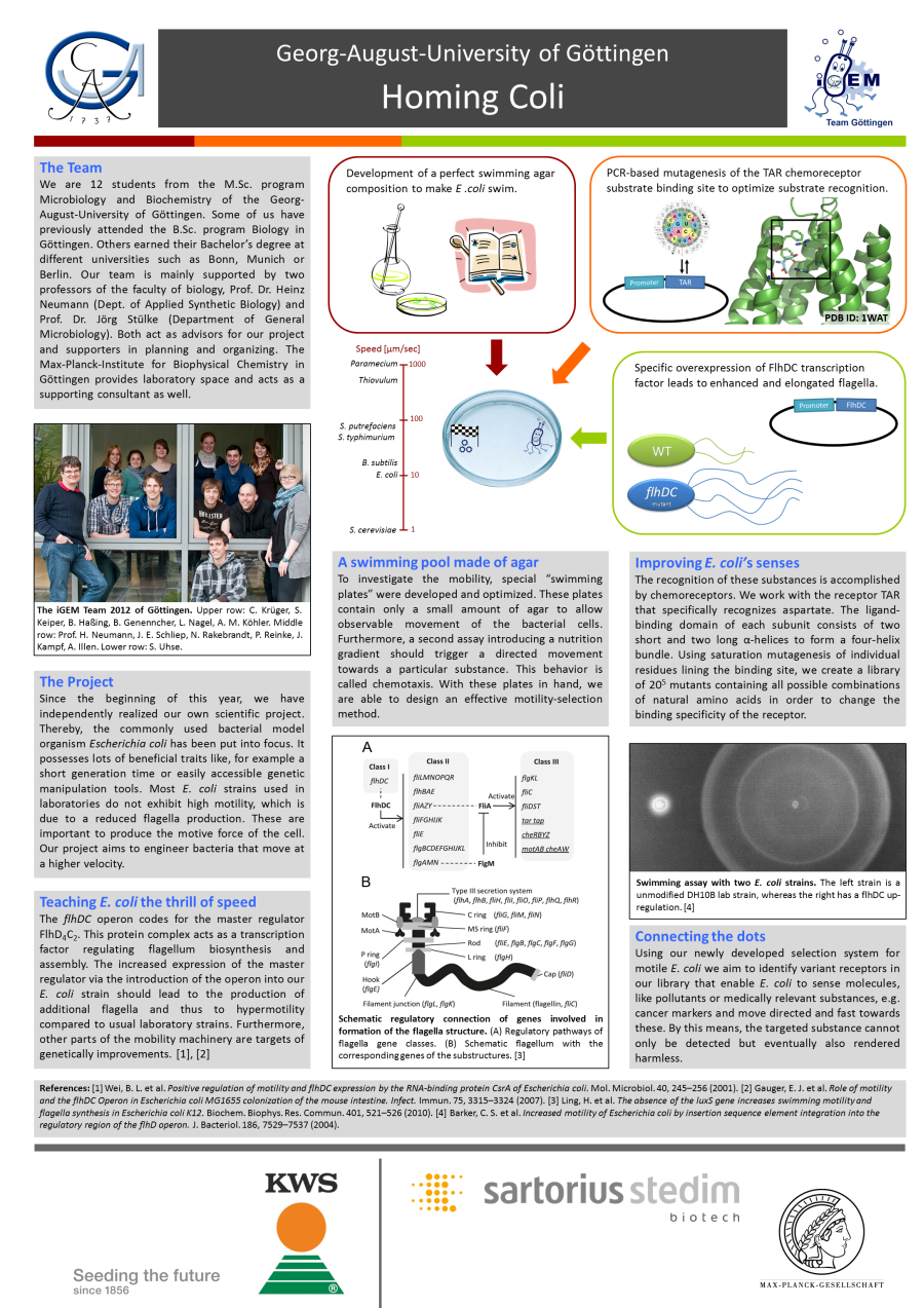
|
↑ Return to top
Team Göttingen Sponsors and Supporter |
 |  |



|
 "
"


