Team:Cambridge/Lab book/Week 6
From 2012.igem.org
(Difference between revisions)
(→Monday) |
(→Friday) |
||
| Line 101: | Line 101: | ||
===Friday=== | ===Friday=== | ||
| + | |||
| + | '''[[Team:Cambridge/Protocols/PCRProtocol|PCR of split Mg2+ vector]]''' | ||
| + | |||
| + | [[File:split Mg2+ 1.jpg|right|250px|thumb|results of split magnesium vector PCR. None of the products worked]] | ||
| + | |||
| + | *PCR of magnesium riboswitch vector repeated with primers to split plasmid (kindly provided by [[Team:Cambridge/Special_thanks|P.J.Steiner]]). | ||
| + | |||
| + | *Lanes 2 + 3: Fragment A (center - cut site (promotor side)) | ||
| + | |||
| + | *Lanes 4 + 5: Fragment B (without 8 codon substitution) (cut site (lac I side) - center) | ||
| + | |||
| + | *Lanes 6 + 7: Fragment B (with 8 codon substitution) (cut stie (lac I side) - center) | ||
| + | |||
| + | *Gels run, found PCR was unsuccessful. | ||
{{Template:Team:Cambridge/CAM_2012_TEMPLATE_FOOT}} | {{Template:Team:Cambridge/CAM_2012_TEMPLATE_FOOT}} | ||
Revision as of 12:09, 6 August 2012
| Week: | 3 | 4 | 5 | 6 | 7 |
|---|
Contents |
Monday
- Not all products were obtained during Friday's PCR. Most of these missing products were large vector backbones. They are being run again, with a much longer extension time of 300s. If that fails, primers will be ordered to split the vectors into manageable chunks, and the PCR reattempted when they arrive.
- PCR cycle x35:
- 15s Denaturing at 95 C
- 45s Annealing at 60 C
- 300s Extension at 72 C
- Remaining stray product had a slightly tricky secondary structure at the 3' end. It will be run at a series of annealing temperatures in a PCR machine capable of a temperature gradient.
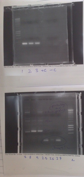
- mOrange PCR run at many different temperatures, from 62 °C to 76 °C. Hopefully this should resolve the difficulties we have been having.
Separation of vector and mOrange DNA
- Positive control produced no band. No primer smear - primers may not be in mix for some reason.
- Realized correct lux vector template was not added during PCR preparation, consequently no amplification occured. Still appears to be a primer smear.
- Fluorescent construct produced several bands. Appears to be due to mis-priming during PCR. Either changing the primers or raising the annealing temperature should solve this problem, but may mean that we have to do this PCR separately.
- Riboswitch vector also failed. However, the extraction from the previous PCR run may have worked. We will try producing a functional plasmid with this extraction before running this PCR again.
Construction of riboswitch plasmid with Gibson Assembly
- DNA from lanes 27+28, 27+29, 27+30 from gels run on Friday fused together with Gibson assembly to produce riboswitch construct. This does not have replacement of the first 8 codons of lac I with the 8 codons native to the gene downstream of the riboswitch.
- DNA from lanes 22,23 and 24 fused with riboswitch DNA produced two weeks ago to produce riboswitch construct. This has replacement of the first 8 codons of lac I with the 8 codons native to the gene downstream of the riboswitch.
Transformation of Bacillus with riboswitch construct
- Plasmids made by Gibson transformed into bacillus cells made two weeks ago and transformants plated out on 5μg/ml chloramphenicol plates.
Tuesday
New biobricks
- E.coli containing biobricks ordered were plated out on kanomycin plates (50μg/ml).
Transformation of Bacillus with riboswitch construct
- Plasmids made by Gibson transformed into bacillus cells made two weeks ago and transformants plated out on 5μg/ml chloramphenicol plates.
Transformation of E.coli with riboswitch construct
- Plasmids made by Gibson transformed into TOP10 e.coli cells and transformants plated out on 100μg/ml ampicillin plates.
Wednesday
Mg2+ Riboswitch
- Successful colonies produced from transformations two days ago streaked out onto chloramphenicol (5 μg/ml) containing plates.
- Colonies also grown up in 10ml of medium A for use with plate reader later.
- Standard PCR rerun, this time splitting fluorescent vector into two separate sections, one of 3kb and one of 4.5kb.
- New primers used:
- Fragment 1 reverse:
- Fragment 2 forward:
Thursday
Friday
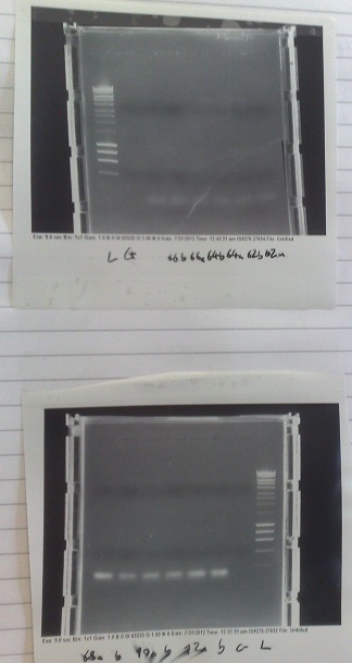
- PCR of magnesium riboswitch vector repeated with primers to split plasmid (kindly provided by P.J.Steiner).
- Lanes 2 + 3: Fragment A (center - cut site (promotor side))
- Lanes 4 + 5: Fragment B (without 8 codon substitution) (cut site (lac I side) - center)
- Lanes 6 + 7: Fragment B (with 8 codon substitution) (cut stie (lac I side) - center)
- Gels run, found PCR was unsuccessful.
 "
"


