Team:UANL Mty-Mexico/Project/recovery
From 2012.igem.org
| Line 7: | Line 7: | ||
<p><b>L2 ribosomal protein</b></p> | <p><b>L2 ribosomal protein</b></p> | ||
<p><br></p> | <p><br></p> | ||
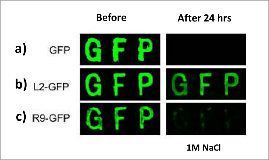
| - | <p>Taniguchi<i> et al.</i> reported in 2007 that the L2 ribosomal protein from <i>E. coli </i>strongly adsorbs to silica surfaces, up to 200 times tighter than poliarginine tags commonly used for protein purification. In their work, Taniguchi <i>et al. </i>constructed a fusion protein of L2 and green fluorescent protein (GFP) which adsorbed to a silica surface even after washing for 24 hours with a buffer containing 1 M NaCl (Figure 1).</p> | + | <p>Taniguchi<i> et al.</i> reported in 2007 that the L2 ribosomal protein from <i>E. coli </i>strongly adsorbs to silica surfaces, up to 200 times tighter than poliarginine tags commonly used for protein purification. In their work, Taniguchi <i>et al. </i> 2007, constructed a fusion protein of L2 and green fluorescent protein (GFP) which adsorbed to a silica surface even after washing for 24 hours with a buffer containing 1 M NaCl (Figure 1).</p> |
<p><br></p> | <p><br></p> | ||
<table class="image" align="center"> | <table class="image" align="center"> | ||
Revision as of 22:27, 26 September 2012
Silica adsorption.
A variety of biorremediation projects have been developed in the last few years; unfortunately none of them has scalable potential due to a lack of an efficient cell recovery system. Therefore developing scalable tools is a very important part of biological systems that could substitute chemical and physical traditional methods that are usually aggressive and environmentally unfriendly (Malik 2004).
L2 ribosomal protein
Taniguchi et al. reported in 2007 that the L2 ribosomal protein from E. coli strongly adsorbs to silica surfaces, up to 200 times tighter than poliarginine tags commonly used for protein purification. In their work, Taniguchi et al. 2007, constructed a fusion protein of L2 and green fluorescent protein (GFP) which adsorbed to a silica surface even after washing for 24 hours with a buffer containing 1 M NaCl (Figure 1).
 |
They mapped the silica-binding domains to amino acids 1-60 and 203-273.
Silica-binding of L2 fusion proteins does not require chemical modifications or pretreatments as opposed to most adsorption methods. Thus developing a cellular silica-binding system seems to have a recovery and scalable potential suited for biorremediation applications.
Expression of L2 in the outer membrane
Even though there are many available carrier proteins to express passengers in E. coli's cell membrane, these systems are mostly designed for small peptides and proteins. On the other hand, membrane proteins AIDA-I and Lpp-OmpA allow the expression of larger proteins in the outer membrane.
AIDA-I is an E. coli membrane protein with a passenger domain of 76 kDa exposed to the extracellular space and a transmembrane beta-barrel domain of 45 kDa; the latter has been used to express functional proteins in the cell-membrane of up to 65 kDa (van Bloois et al. 2011). Furthermore passengers coupled to AIDA-I have been reported to reach an expression level of more than 100 000 copies per cell in the outer membrane (Jose & Meyer 2007), therefore making this expression system a great option for the expression of L2 in E. coli's outer membrane (Figure 2a, Li et al. 2007).
 |
Another carrier protein is Lpp-OmpA, which is a fusion protein of two E. coli's native proteins: signal-peptide from Lpp lipoprotein and OmpA transmembrane protein (Francisco et al., 1992). This system has been used to carry proteins from 27 up to 74 kDa and reaches an expression level of up to 100,000 copies per cell in the outer membrane (van Bloois et al., 2011), although the structure is different from AIDA-I (figure 2b). Figure 3 represents the fusion proteins arrangement in the outer membrane of E. coli.
 |
 "
"
