 "
"
Team:UC Chile/Bactomithril
From 2012.igem.org
| Line 63: | Line 63: | ||
== Results== | == Results== | ||
| - | < | + | You can see detailed informarion of our results in <a href="https://2012.igem.org/Team:UC_Chile/bactoresults">this section </a>. Nevertheless, we can briefly summarize the following: we know that sfGFP expresses in our e. coli, but we don’t know if it is attached to the external side of the membrane, or if the HIV-1 protease works as expected. As this secretion system seems to be failing, we chose to try other alternatives. |
| - | + | == Future Prospects: The construction of the first spider silk biobrick == | |
| - | + | ||
| - | + | ||
| - | + | ||
| - | + | ||
| - | + | ||
| - | + | ||
| - | + | ||
| - | + | ||
| - | + | ||
| - | + | ||
| - | + | ||
| - | + | ||
| - | + | ||
| - | + | ||
| - | + | ||
| - | + | ||
| - | + | ||
| - | + | ||
| - | + | ||
| - | + | ||
| - | + | ||
| - | + | ||
| - | + | ||
| - | + | ||
| - | + | ||
| - | + | ||
| - | + | ||
| - | + | ||
| - | + | ||
| - | + | ||
| - | + | ||
| - | + | ||
| - | + | ||
| - | + | ||
| - | + | ||
| - | + | ||
| - | + | ||
| - | + | ||
| - | + | ||
| - | + | ||
| - | + | ||
| - | + | ||
| - | + | ||
| - | + | ||
| - | + | ||
| - | + | ||
| - | + | ||
| - | + | ||
| - | + | ||
| - | + | ||
| - | + | ||
| - | + | ||
| - | + | ||
| - | + | ||
| - | + | ||
| - | + | ||
| - | + | ||
| - | + | ||
| - | + | ||
| - | + | ||
| - | + | ||
| - | + | ||
| - | + | ||
| - | + | ||
| - | + | ||
| - | + | ||
| - | + | ||
| - | + | ||
| - | + | ||
| - | + | ||
| - | + | ||
| - | + | ||
| - | + | ||
| - | + | ||
| - | + | ||
| - | + | ||
| - | + | ||
| - | + | ||
| - | + | ||
| - | + | ||
| - | + | ||
| - | + | ||
| - | + | ||
| - | + | ||
| - | + | ||
| - | + | ||
| - | + | ||
| - | + | ||
| - | + | ||
| - | + | ||
| - | + | ||
We tried with a lot of patience (in SEVERAL attempts with many different strategies) to make a biobrick with the spider silk monomer ADF-3 or the sequence of DNA corresponding to ADF-3 and tags necessary to identify and export the protein with the Type 3 Secretion System of Salmonella (SPI 2). The original sequences are described in | We tried with a lot of patience (in SEVERAL attempts with many different strategies) to make a biobrick with the spider silk monomer ADF-3 or the sequence of DNA corresponding to ADF-3 and tags necessary to identify and export the protein with the Type 3 Secretion System of Salmonella (SPI 2). The original sequences are described in | ||
Revision as of 23:49, 23 October 2012

Contents |
Overview
Spider dragline silk is an exceptionally strong and elastic fiber and is an ideal material for numerous biomedical and industrial applications. In this secondary project we developed a novel secretion system that allows to export proteins to the extracellular medium and tried to add the first spider-silk biobrick to the Registry. This biobrick would have the synthetic silk gene ADF-3 from the orb weaving spider Araneus diadematus. Nevertheless, due to the repetitive nature of the spider silk monomer we could not finish this biobrick for the Regional Jamboree (despite several attempts and strategies).
Animation describing the first secretion system

.gif)
Introduction
Spider dragline silk is an exceptionally strong and elastic biomaterial and one of the strongest natural fibers known. It is five times stronger by weight than steel and three times tougher than the top quality man-made fiber Kevlar [1]. That is why it is referred to as “biosteel”. Furthermore, this material is biocompatible, biodegradable and has potential for processing in aqueous solution under ambient conditions.
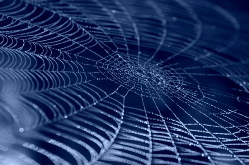
These properties make the spider silk an ideal material for numerous industrial applications such as bullet-proof vests, parachutes, seat belts and composite materials in aircraft, and a promising substance for biomedical applications such as drug delivery systems and scaffolds for tissue engineering.
The excellent mechanical properties and low immunogenicity of spider webs have fascinated the man for thousands of years, but unfortunately large scale farming of spiders is not convenient because of the highly territorial and aggressive character of these arthropods. Synthetic spider silk production is a promising way to make use of this exceptional biomaterial.
Construct: a novel secretion system
Working Plasmid (BBa_K43011)

GFP Reporter Plasmid (BBa_K43012)

Protease Producer Plasmid (BBa_K43013)

Notebook
In this section you will find the complete description of our work in this project.
Results
You can see detailed informarion of our results in <a href="https://2012.igem.org/Team:UC_Chile/bactoresults">this section </a>. Nevertheless, we can briefly summarize the following: we know that sfGFP expresses in our e. coli, but we don’t know if it is attached to the external side of the membrane, or if the HIV-1 protease works as expected. As this secretion system seems to be failing, we chose to try other alternatives.
Future Prospects: The construction of the first spider silk biobrick
We tried with a lot of patience (in SEVERAL attempts with many different strategies) to make a biobrick with the spider silk monomer ADF-3 or the sequence of DNA corresponding to ADF-3 and tags necessary to identify and export the protein with the Type 3 Secretion System of Salmonella (SPI 2). The original sequences are described in
Widmaier, D. M., Tullman-Ercek, D., Mirsky, E. a, Hill, R., Govindarajan, S., Minshull, J., & Voigt, C. a. (2009). Engineering the Salmonella type III secretion system to export spider silk monomers. Molecular systems biology, 5(309), 309. doi:10.1038/msb.2009.62
This sequence was kindly sent by Dr. Christopher Voigt to us.
A detailed description of this work is in the notebook section. After tons of hours of hard work, when we finally obtained PCR amplified bands of the correct size, the digestion tests were positive, colony PCR were succesful and Gibson Assemblies seemed to work, we sent our final construct for sequencing and the results did not match with our designs. Probably this is due to the hihgly repetitive nature of the spider silk monomers.
References
In this section you will find references about production and modelling of recombinant spider silk proteins.
Recommended literature
Teulé, F., Cooper, A. R., Furin, W. a, Bittencourt, D., Rech, E. L., Brooks, A., & Lewis, R. V. (2009). A protocol for the production of recombinant spider silk-like proteins for artificial fiber spinning. Nature protocols, 4(3), 341-55. doi:10.1038/nprot.2008.250
Widhe, M., Johansson, J., Hedhammar, M., & Rising, A. (2012). Invited review current progress and limitations of spider silk for biomedical applications. Biopolymers, 97(6), 468-78. doi:10.1002/bip.21715
Widmaier, D. M., Tullman-Ercek, D., Mirsky, E. a, Hill, R., Govindarajan, S., Minshull, J., & Voigt, C. a. (2009). Engineering the Salmonella type III secretion system to export spider silk monomers. Molecular systems biology, 5(309), 309. doi:10.1038/msb.2009.62
Xia, X.-X., Qian, Z.-G., Ki, C. S., Park, Y. H., Kaplan, D. L., & Lee, S. Y. (2010). Native-sized recombinant spider silk protein produced in metabolically engineered Escherichia coli results in a strong fiber. Proceedings of the National Academy of Sciences of the United States of America, 107(32), 14059-63. doi:10.1073/pnas.1003366107
Complementary literature
An, B., Hinman, M. B., Holland, G. P., Yarger, J. L., & Lewis, R. V. (2011). Inducing β-sheets formation in synthetic spider silk fibers by aqueous post-spin stretching. Biomacromolecules, 12(6), 2375-81. doi:10.1021/bm200463e
Askarieh, G., Hedhammar, M., Nordling, K., Saenz, A., Casals, C., Rising, A., Johansson, J., et al. (2010). Self-assembly of spider silk proteins is controlled by a pH-sensitive relay. Nature, 465(7295), 236-8. doi:10.1038/nature08962
Ayoub, N. a, Garb, J. E., Tinghitella, R. M., Collin, M. a, & Hayashi, C. Y. (2007). Blueprint for a high-performance biomaterial: full-length spider dragline silk genes. PloS one, 2(6), e514. doi:10.1371/journal.pone.0000514
Bauer, F., & Scheibel, T. (2012). Artificial egg stalks made of a recombinantly produced lacewing silk protein. Angewandte Chemie (International ed. in English), 51(26), 6521-4. doi:10.1002/anie.201200591
Bayley, H. (1998). Puri ® cation and characterization of recombinant spider silk expressed in Escherichia coli. Cell, 1, 31-38.
Bogush, V. G., Sokolova, O. S., Davydova, L. I., Klinov, D. V., Sidoruk, K. V., Esipova, N. G., Neretina, T. V., et al. (2009). A novel model system for design of biomaterials based on recombinant analogs of spider silk proteins. Journal of neuroimmune pharmacology : the official journal of the Society on NeuroImmune Pharmacology, 4(1), 17-27. doi:10.1007/s11481-008-9129-z
Dams-Kozlowska, H., Majer, A., Tomasiewicz, P., Lozinska, J., Kaplan, D. L., & Mackiewicz, A. (2012). Purification and cytotoxicity of tag-free bioengineered spider silk proteins. Journal of biomedical materials research. Part A, 1-9. doi:10.1002/jbm.a.34353
Das, R., & Baker, D. (2008). Macromolecular modeling with rosetta. Annual review of biochemistry, 77, 363-82. doi:10.1146/annurev.biochem.77.062906.171838
Kenney, J. M., Knight, D., Wise, M. J., & Vollrath, F. (2002). Amyloidogenic nature of spider silk. European Journal of Biochemistry, 269(16), 4159-4163. doi:10.1046/j.1432-1033.2002.03112.x
Keten, S., & Buehler, M. J. (2010). Nanostructure and molecular mechanics of spider dragline silk protein assemblies. Journal of the Royal Society, Interface / the Royal Society, 7(53), 1709-21. doi:10.1098/rsif.2010.0149
Lazaris, A., Arcidiacono, S., Huang, Y., Zhou, J.-F., Duguay, F., Chretien, N., Welsh, E. a, et al. (2002). Spider silk fibers spun from soluble recombinant silk produced in mammalian cells. Science (New York, N.Y.), 295(5554), 472-6. doi:10.1126/science.1065780
Moisenovich, M. M., Pustovalova, O. L., Arhipova, a Y., Vasiljeva, T. V., Sokolova, O. S., Bogush, V. G., Debabov, V. G., et al. (2011). In vitro and in vivo biocompatibility studies of a recombinant analogue of spidroin 1 scaffolds. Journal of biomedical materials research. Part A, 96(1), 125-31. doi:10.1002/jbm.a.32968
Rising, A., Widhe, M., Johansson, J., & Hedhammar, M. (2011). Spider silk proteins: recent advances in recombinant production, structure-function relationships and biomedical applications. Cellular and molecular life sciences : CMLS, 68(2), 169-84. doi:10.1007/s00018-010-0462-z
Tobar, J. a, Carreño, L. J., Bueno, S. M., González, P. a, Mora, J. E., Quezada, S. a, & Kalergis, A. M. (2006). Virulent Salmonella enterica serovar typhimurium evades adaptive immunity by preventing dendritic cells from activating T cells. Infection and immunity, 74(11), 6438-48. doi:10.1128/IAI.00063-06
Vollrath, F. (1999). Biology of spider silk. International journal of biological macromolecules, 24(2-3), 81-8. Retrieved from http://www.ncbi.nlm.nih.gov/pubmed/10342751
Vollrath, F., & Knight, D. P. (2001). Liquid crystalline spinning of spider silk. Nature, 410(6828), 541-8. doi:10.1038/35069000
Winkler, S., Szela, S., Avtges, P., Valluzzi, R., Kirschner, D. a, & Kaplan, D. (1999). Designing recombinant spider silk proteins to control assembly. International journal of biological macromolecules, 24(2-3), 265-70. Retrieved from http://www.ncbi.nlm.nih.gov/pubmed/10342773


