|
|
| Line 1: |
Line 1: |
| | <html></p></html>{{Template:Peking2012_Color_Prologue}}{{Template:Peking2012_Color_Modeling}}<html> | | <html></p></html>{{Template:Peking2012_Color_Prologue}}{{Template:Peking2012_Color_Modeling}}<html> |
| | <script type="text/javascript"> | | <script type="text/javascript"> |
| - | sublists_Now = 0; | + | sublists_Now = 1; |
| | var subsubitem=subfirst.getElementsByTagName('ul')[sublists_Now].getElementsByTagName('a')[0]; | | var subsubitem=subfirst.getElementsByTagName('ul')[sublists_Now].getElementsByTagName('a')[0]; |
| | subsubitem.style.color='#60b0f0'; | | subsubitem.style.color='#60b0f0'; |
| Line 8: |
Line 8: |
| | </script> | | </script> |
| | <div class="PKU_context floatR first"> | | <div class="PKU_context floatR first"> |
| - | <h3 id="title1">Summary</h3> | + | <h3 id="title2">Network System</h3> |
| | <p> | | <p> |
| - | Our <i>Luminesensor</i> is a fusion protein to sense 450nm to 470nm light and then regulate the gene expression.(<a href="/Team:Peking/Project/Luminesensor/Future#FigS">spectrum data here</a>) Although Luminesensor excels and eclipses similar systems due to its ultra-sensitivity and dynamic range, there are still several imperfect aspects. For example, the response time of the protein can be up to hours<sup><a href="#ref1" title="Light Activation of the LOV Protein Vivid Generates a Rapidly Exchanging Dimer. B. D. Zoltowski etc. Biochemistry">[1]</a></sup> and the contrast of binding efficiency with and without light has much room for improvement. After modeling the DNA binding process of <i>Luminesensor</i>, we managed to find out four key parameters, two of which mainly control the response time, and the others control the contrast of binding efficiency. According to the feasibility in experiment, we tuned two most facile parameters of each aspect to optimize the performance of <i>Luminsensor</i>, and later figured out mutation sites related to these two parameters experimentally.
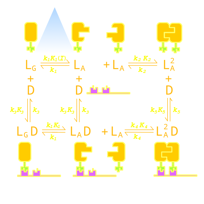
| + | Firstly we established a reaction network for the DNA binding process of <i>Luminesensor</i>. In order to quantify this system, we used <a href="/Team:Peking/Modeling/Appendix/ODE">ODE (Oridinary Differential Equations)</a> model at the beginning. Previous works by other scientists indicate that the Vivid (VVD) protein, a sensing domain of <i>Luminesensor</i>, dimerizes in the presence of light,<sup><a href="#ref1" title="Light Activation of the LOV Protein Vivid Generates a Rapidly Exchanging Dimer. B. D. Zoltowski etc. Biochemistry">[1]</a></sup> and the LexA protein, a binding domain of <i>Luminesensor</i>, binds at specific sequences on DNA predominantly when coupled<sup><a href="#ref2" title="LexA Repressor Forms Stable Dimers in Solution. R. Mohana-Borges etc. THE JOURNAL OF BIOLOGICAL CHEMISTRY">[2]</a></sup> and subsequently represses the interest gene. By combining with the mechanisms of the two functional domains of Luminesensor, we concluded with the following network: |
| - | </p>
| + | |
| - | </div>
| + | |
| - | <div class="PKU_context floatR">
| + | |
| - | <h3 id="title2">Network System with ODE Model</h3>
| + | |
| - | <p>
| + | |
| - | Firstly we established a reaction network for the DNA binding process of <i>Luminesensor</i>. In order to quantify this system, we used <a href="/Team:Peking/Modeling/Background/ODE">ODE (Oridinary Differential Equations)</a> model at the beginning. Previous works by other scientists indicate that the Vivid (VVD) protein, a sensing domain of <i>Luminesensor</i>, dimerizes in the presence of light,<sup><a href="#ref1" title="Light Activation of the LOV Protein Vivid Generates a Rapidly Exchanging Dimer. B. D. Zoltowski etc. Biochemistry">[1]</a></sup> and the LexA protein, a binding domain of <i>Luminesensor</i>, binds at specific sequences on DNA predominantly when coupled<sup><a href="#ref2" title="LexA Repressor Forms Stable Dimers in Solution. R. Mohana-Borges etc. THE JOURNAL OF BIOLOGICAL CHEMISTRY">[2]</a></sup> and subsequently represses the interest gene. By combining with the mechanisms of the two functional domains of Luminesensor, we concluded with the following network: | + | |
| | </p> | | </p> |
| | <div class="floatC"> | | <div class="floatC"> |
| - | [fig 1: Reaction Network (Cartoon Style)]
| + | <img src="/wiki/images/3/3a/Peking2012_LuminesensorNodes.png" alt="Network System Fig" /> |
| - | <p class="description">Fig 1. </p> | + | <p class="description">Fig 1. Network System of our Luminesensor</p> |
| | </div> | | </div> |
| | <p>where</p><ul><li> | | <p>where</p><ul><li> |
| - | L denotes Luminesensor,</li><li> | + | L : Luminesensor,</li><li> |
| - | L<sub>G</sub> denotes the ground state --- with its VVD N-cap locked,</li><li> | + | L<sub>G</sub> : the ground state --- with its VVD N-cap locked,</li><li> |
| - | L<sub>A</sub> denotes the active state --- with its VVD N-cap released,</li><li> | + | L<sub>A</sub> : the active state --- with its VVD N-cap released,</li><li> |
| - | D<sub>L</sub> denotes the specific DNA binding site to Luminesensor.</li></ul> | + | D<sub>L</sub> : the specific DNA binding site to <i>Luminesensor</i>.</li></ul> |
| - | <p>
| + | |
| - | Since no multi-intermediate reactions are hidden in the network above, all reactions can be regarded as elementary reactions. We list all the differential equations as following:
| + | |
| - | </p>
| + | |
| - | <div class="floatC">
| + | |
| - | [fig 2: Ordinary Equations]
| + | |
| - | <p class="description">Fig 2. </p>
| + | |
| - | </div>
| + | |
| - | <p>with parameters listed following:</p>
| + | |
| - | <div class="floatC">
| + | |
| - | <table>
| + | |
| - | <tr>
| + | |
| - | <td>Parameter</td><td>Value</td><td>Unit</td><td>Description</td><td>Source</td>
| + | |
| - | </tr><tr>
| + | |
| - | <td>k<sub>1</sub></td><td>3.x10<sup>-4</sup></td><td>s<sup>-1</sup></td><td>vivid decay rate constant</td><td></td>
| + | |
| - | </tr><tr>
| + | |
| - | <td>k<sub>2</sub></td><td>5.6x10<sup>-5</sup></td><td>s<sup>-1</sup></td><td>vivid dissociation rate constant</td><td><a href="#ref3" title="Mechanism-based tuning of a LOV domain photoreceptor, Brian D. Zoltowski, etc. NATURE CHEMICAL BIOLOGY">[3]</a></td>
| + | |
| - | </tr><tr>
| + | |
| - | <td>k<sub>3</sub></td><td>8.x10<sup>-4</sup></td><td>s<sup>-1</sup></td><td>monomer LexA releasing rate constant from specific binding site</td><td></td>
| + | |
| - | </tr><tr>
| + | |
| - | <td>k<sub>4</sub></td><td>1.x10<sup>-3</sup></td><td>s<sup>-1</sup></td><td>binded monomer LexA dissociation rate constant</td><td></td>
| + | |
| - | </tr><tr>
| + | |
| - | <td>k<sub>5</sub></td><td>1.x10<sup>-4</sup></td><td>s<sup>-1</sup></td><td>dimered LexA releasing rate constant from specific binding site</td><td></td>
| + | |
| - | </tr><tr>
| + | |
| - | <td>K<sub>1</sub>(Dark)</td><td>0</td><td>1</td><td>equilibrium excitation constant on dark</td><td></td>
| + | |
| - | </tr><tr>
| + | |
| - | <td>K<sub>1</sub>(Light)</td><td>1.x10<sup>+3</sup></td><td>1</td><td>equilibrium excitation constant on light</td><td></td>
| + | |
| - | </tr><tr>
| + | |
| - | <td>K<sub>2</sub></td><td>7.7x10<sup>-5</sup></td><td>(n mol/L)<sup>-1</sup></td><td>vivid association equilibrium constant</td><td><a href="#ref4" title="Protein Vivid Generates a Rapidly Exchanging Dimer, Brian D. Zoltowski, etc. BIOCHEMISTRY">[4]</a></td>
| + | |
| - | </tr><tr>
| + | |
| - | <td>K<sub>3</sub></td><td>1.x10<sup>-3</sup></td><td>(n mol/L)<sup>-1</sup></td><td>monomer LexA binding equilibrium constant with specific binding site</td><td><a href="#ref2" title="LexA Repressor Forms Stable Dimers in Solution, R.Mohana-Borges, etc. THE JOURNAL OF BIOLOGICAL CHEMISTRY">[2]</a></td>
| + | |
| - | </tr><tr>
| + | |
| - | <td>K<sub>4</sub></td><td>K<sub>2</sub>xK<sub>5</sub>/K<sub>3</sub></td><td>(n mol/L)<sup>-1</sup></td><td>binded monomer LexA association equilibrium constant</td><td>Thermal Principle</td>
| + | |
| - | </tr><tr>
| + | |
| - | <td>K<sub>5</sub></td><td>1.</td><td>(n mol/L)<sup>-1</sup></td><td>dimered LexA binding equilibrium constant</td><td><a href="#ref2" title="LexA Repressor Forms Stable Dimers in Solution, R.Mohana-Borges, etc. THE JOURNAL OF BIOLOGICAL CHEMISTRY">[2]</a></td>
| + | |
| - | </tr><tr>
| + | |
| - | <td>[L<sub>G</sub>]<sub>0</sub></td><td>1000</td><td>n mol/L</td><td>initial concentration of Luminesensor in ground state</td><td></td>
| + | |
| - | </tr><tr>
| + | |
| - | <td>[L<sub>A</sub>]<sub>0</sub></td><td>0</td><td>n mol/L</td><td>initial concentration of Luminesensor in active state</td><td></td>
| + | |
| - | </tr><tr>
| + | |
| - | <td>[L<sub>A</sub><sup>2</sup>]<sub>0</sub></td><td>0</td><td>n mol/L</td><td>initial concentration of dimered Luminesensor</td><td></td>
| + | |
| - | </tr><tr>
| + | |
| - | <td>[D<sub>L</sub>]<sub>0</sub></td><td>100</td><td>n mol/L</td><td>initial concentration of free specific binding site on DNA</td><td>high-copy plasmid</td>
| + | |
| - | </tr><tr>
| + | |
| - | <td>[L<sub>G</sub>D<sub>L</sub>]<sub>0</sub></td><td>0</td><td>n mol/L</td><td>initial concentration of dimered Luminesensor binded Luminesensor in ground state</td><td></td>
| + | |
| - | </tr><tr>
| + | |
| - | <td>[L<sub>A</sub>D<sub>L</sub>]<sub>0</sub></td><td>0</td><td>n mol/L</td><td>initial concentration of dimered Luminesensor binded Luminesensor in active state</td><td></td>
| + | |
| - | </tr><tr>
| + | |
| - | <td>[L<sub>A</sub><sup>2</sup>D<sub>L</sub>]<sub>0</sub></td><td>0</td><td>n mol/L</td><td>initial concentration of binded and dimered Luminesensor</td><td></td>
| + | |
| - | </tr>
| + | |
| - | </table>
| + | |
| - | <p class="description">Tab 1. Reaction Parameters</p>
| + | |
| - | </div>
| + | |
| - | <p>and then simulated the dynamic behavior of the components with our result shown below:</p>
| + | |
| - | <div class="floatC">
| + | |
| - | [fig 3: ODE Simulation]
| + | |
| - | <p class="description">Fig 3. Simulation of Origin System</p>
| + | |
| - | </div>
| + | |
| - | <div>
| + | |
| - | <p>where the binding rate</p><p style="text-align:center;">
| + | |
| - | r<sub>b</sub> = 1 - [D<sub>L</sub>]/[D<sub>T</sub>]
| + | |
| - | <br />(D<sub>T</sub> means the total concentration of specific binding site on DNA)
| + | |
| - | </p><p>indicates the repressing degree, and the dimerizing rate</p><p style="text-align:center;">
| + | |
| - | r<sub>d</sub> = 2[L<sub>A</sub><sup>2</sup>X]/[L<sub>T</sub>]
| + | |
| - | <br />(L<sub>T</sub> means the total concentration of Luminesensor, X denotes D<sub>L</sub> or nothing)
| + | |
| - | </p><p>indicates the dimerizing degree, and the activating rate</p><p style="text-align:center;">
| + | |
| - | r<sub>a</sub> = ([L<sub>A</sub>X] + 2[L<sub>A</sub><sup>2</sup>X])/[L<sub>T</sub>]
| + | |
| - | </p>
| + | |
| - | </div>
| + | |
| - | <div>
| + | |
| - | <p>
| + | |
| - | From the figure above, we discovered that the activation and decay of Luminesensor are the pioneers of progress, and the activating rate is the most completely switched variable as lighting varies. The promoter sequences in the DNA are repressed even though the <i>Luminesensor</i> has not thoroughly dimered.
| + | |
| - | </p>
| + | |
| - | </div>
| + | |
| - | </div>
| + | |
| - | <div class="PKU_context floatR">
| + | |
| - | <h3 id="title3">Stochastic Simulation</h3>
| + | |
| - | <p>
| + | |
| - | In order to check the working stability of Luminesensor, we simulated this reaction network with a <a href="/Team:Peking/Modeling/Background/Stochastic">stochastic model</a>. By estimating the volume of a cell, we converted the concentration of a component into the number of molecules by 1 n mol/L : 1. The result are shown below:
| + | |
| - | </p>
| + | |
| - | <div class="floatC">
| + | |
| - | [fig 4: Stochastic Result]
| + | |
| - | <p class="description">Fig 4. Stochastic Simulation</p>
| + | |
| - | </div>
| + | |
| - | <p>
| + | |
| - | According to the figure above, the noise did not influence this system. Thus, the <i>Luminesensor</i> is expected to work theoretically. Besides, the average value of stochastic simulation is coupled with the result of ODE model, which in turn proves the self-consistency of our ODE model.
| + | |
| - | </p>
| + | |
| - | </div>
| + | |
| - | | + | |
| - | | + | |
| - | <div class="PKU_context floatR">
| + | |
| - | <h3 id="title4">Parameter Analysis & Optimization</h3>
| + | |
| - | <p>
| + | |
| - | After modeling the origin system, we attempted to optimize it in a rational way. We have tuned the parameters both up and down, one by one, and finally discovered four parameters which predominantly influence this system.
| + | |
| - | </p><ul>
| + | |
| - | <li>k1 (Vivid decay rate constant)</li>
| + | |
| - | <li>k3 (rate constant of monomer LexA releasing from specific binding site)</li>
| + | |
| - | </ul><p>determines the time scale.</p><ul>
| + | |
| - | <li>K2 (Vivid association equilibrium constant)</li>
| + | |
| - | <li>K5 (dimered LexA binding equilibrium constant)</li>
| + | |
| - | </ul><p>determine the contrast between dark and light. Here shows the simulation data:
| + | |
| - | </p>
| + | |
| - | <div class="floatC">
| + | |
| - | [fig 5: Parameter Tuning]
| + | |
| - | <p class="description">Fig 5. Parameter Tuning, the left shows the simulation by ODE model and the right shows the stochastic simulation. (a) the origin system. (b) tuning k1 up by 10 times while K1 remains unchanged. (c) tuning k3 up by 10 times while K3 remains unchanged. (d) tuning K2 up by 10 times. (e) tuning K5 up by 10 times.</p>
| + | |
| - | </div>
| + | |
| - | <p>
| + | |
| - | As for contrast optimizing, we define an expression
| + | |
| - | </p><p style="text-align:center;">
| + | |
| - | completely-repressing rate = completely-repressing time / total sample time
| + | |
| - | </p><p>
| + | |
| - | as the interest, where completely-repressing means that all specific binding sites are occupied by <i>Luminesensor</i>. The higher completely-repressing rate is, the better the Luminesensor represses. The completely-repressing rate of each simulation is shown at the right of Figure 5. Within the two chosen parameters, K2 (vivid association equilibrium constant) is related to the association mechanism of Vivid protein and K5 (dimered LexA binding equilibrium constant) is related to the cooperative binding mechanism.
| + | |
| - | <br /><br />
| + | |
| - | As for speed optimizing, k1 (vivid decay rate constant) is related to the activation mechanism of Vivid protein and k3 (monomer LexA releasing rate constant from specific binding site) is related to the binding mechanism, thus also to the LexA and sequence. If we change the binding affinity of the sequence, then it is difficult to make constant K3 (monomer LexA binding equilibrium constant with specific binding site), whose variance is predicted to ruin the contrast of this system from simulation. Therefore, we chose to apply a mutation on the Vivid protein in order to attain a faster <i>Luminesensor</i>, which has high levels of k1 (vivid decay rate constant).
| + | |
| - | <br /><br />
| + | |
| - | By searching the data of mutant in vivid protein among recent papers, we focus on these mutants: M135I in vivid dimerization domain to enhance K2 (vivid association equilibrium constant)<sup><a href="#ref?" title="">[?]</a></sup> and I74V of amino acids surrounding Cys108 to enhance k1 (vivid decay rate constant)<sup><a href="#ref3" title="Mechanism-based tuning of a LOV domain photoreceptor, Brian D. Zoltowski, etc. NATURE CHEMICAL BIOLOGY">[3]</a></sup>. As for K5 (dimered LexA binding equilibrium constant), we chose LexA408, the mutant of LexA.<sup><a href="#ref5" title="A new LexA-based genetic system for monitoring and analyzing protein heterodimerization in Escherichia coli, M. Dmitrova. etc. Springer-Verlag Mol Gen Genet">[5]</a></sup> However, our main reason for choosing LexA408 over the wild-type LexA is due to the bio-orthogonality between them.
| + | |
| - | </p>
| + | |
| - | </div>
| + | |
| - | <div class="PKU_context floatR">
| + | |
| - | <h3 id="title5">Orthogonal Test <i>in silico</i></h3>
| + | |
| - | <p>
| + | |
| - | To modularize the genetic system, our <i>Luminesensor</i> is expected to be bio-orthogonal with the origin system in bacteria. LexA, a natural element from the lactin-SOS system in bacteria may cause unexpected crosstalk. In order to remove this obstacle on the application prospects of our <i>Luminesensor</i>, we use LexA408 instead of the wild-type LexA. LexA408 and LexA are bio-orthogonal with each other since the sequence of the binding sites have variations.
| + | |
| - | <br /><br />
| + | |
| - | By adding several nodes into the network, we constructed modeling for orthogonality test:
| + | |
| - | </p>
| + | |
| - | <div class="floatC">
| + | |
| - | [fig 6: Reaction Network for Orthogonal Test]
| + | |
| - | <p class="description">Fig 6. Reaction Network for Orthogonal Test</p>
| + | |
| - | </div>
| + | |
| - | <p>where</p><ul><li>
| + | |
| - | L denotes <i>Luminesensor</i></li><li>
| + | |
| - | I denotes the inner wild LexA</li><li>
| + | |
| - | D<sub>L</sub> denotes the specific DNA binding site to <i>Luminesensor</i></li><li>
| + | |
| - | D<sub>I</sub> denotes the specific DNA binding site to wild LexA</li></ul>
| + | |
| - | <p>The parameters are estimated as following:</p>
| + | |
| - | <div class="floatC">
| + | |
| - | <table>
| + | |
| - | <tr>
| + | |
| - | <td>Parameter</td><td>Value</td><td>Unit</td><td>Description</td><td>Source</td>
| + | |
| - | </tr><tr>
| + | |
| - | <td>k<sub>6</sub></td><td>1.x10<sup>-4</sup></td><td>s<sup>-1</sup></td><td>dimered LexA releasing rate constant from non-specific binding site</td><td></td>
| + | |
| - | </tr><tr>
| + | |
| - | <td>K<sub>6</sub></td><td>1.x10<sup>-2</sup></td><td>(n mol/L)<sup>-1</sup></td><td>dimered non-specific binding equilibrium constant</td><td><a href="#ref9">[9]</a></td>
| + | |
| - | </tr>
| + | |
| - | </table>
| + | |
| - | <p class="description">Tab 2. Reaction Parameters for Orthogonal Test</p>
| + | |
| - | </div>
| + | |
| - | <div class="floatC">
| + | |
| - | [fig 7: Orthogonal Test Result]
| + | |
| - | <p class="description">Fig 7. Orthogonal Test Result, the left show the simulation by ODE model and the right shows the stochastic simulation. (a) the origin system. (b) competition induced. (c) competition induced and tuning K6 up by 10 times.
| + | |
| - | </p>
| + | |
| - | </div>
| + | |
| - | <p>
| + | |
| - | The result shows that the endurance of K6/K5 is up to around 1%. Our <i>Luminesensor</i> can be used in bacteria with wild-type LexA.
| + | |
| - | </p>
| + | |
| - | </div>
| + | |
| - | <div class="PKU_context floatR">
| + | |
| - | <h3 id="title6">Conclusion</h3>
| + | |
| | <p> | | <p> |
| - | The modeling above points out a way to optimize our <i>Luminesensor</i> -- the two critical mutation. It also shows the system still works well even if considering noise and inner competition.
| + | Since no multi-intermediate reactions are hidden in the network above, all reactions can be regarded as elementary reactions. |
| | </p> | | </p> |
| - | </div>
| |
| - | <div class="PKU_context floatR">
| |
| - | <h3 id="title7">Referrence</h3>
| |
| - | <p></p>
| |
| - | <ul class="refer"><li id="ref1">
| |
| - | [1] Light Activation of the LOV Protein Vivid Generates a Rapidly Exchanging Dimer. B. D. Zoltowski etc. Biochemistry
| |
| - | </li><li id="ref2">
| |
| - | [2] LexA Repressor Forms Stable Dimers in Solution. R. Mohana-Borges etc. THE JOURNAL OF BIOLOGICAL CHEMISTRY
| |
| - | </li><li id="ref3">
| |
| - | [3] Mechanism-based tuning of a LOV domain photoreceptor, Brian D. Zoltowski, etc. NATURE CHEMICAL BIOLOGY
| |
| - | </li><li id="ref4">
| |
| - | [4] Protein Vivid Generates a Rapidly Exchanging Dimer, Brian D. Zoltowski, etc. BIOCHEMISTRY
| |
| - | </li><li id="ref5">
| |
| - | [5] A new LexA-based genetic system for monitoring and analyzing protein heterodimerization in Escherichia coli, M. Dmitrova. etc. Springer-Verlag Mol Gen Genet
| |
| - | </li></ul>
| |
| | </div> | | </div> |
| | </html>{{Template:Peking2012_Color_Epilogue}} | | </html>{{Template:Peking2012_Color_Epilogue}} |
 "
"