 "
"
Team:Peking/Modeling/Channel
From 2012.igem.org


![]()
Motivation
Light-oxygen-voltage (LOV) domain containing fusion proteins are validated photosensing devices that have been widely utilized and well characterized in the field of optogenetics. The use of Vivid (VVD) in the construction of an ultra-sensitive Luminesensor (LexA-VVD) by Peking iGEM 2012 has opened the gate to a new generation of optogentics.
Functional the existing biobrick may be, there are two major concerns about it:
- 1. Blue light absorption at 455nm (Fig. 1, Dark) suffers from a disability of penetrating tissues for further applications such as phototherapy, because of its short wavelength.
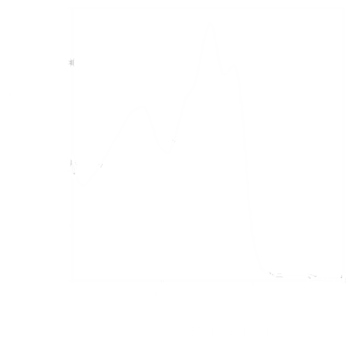
Figure 1. Absorption Spectrum of VVD protein[3]
- 2. Mono-channel manipulation of cellular behavior is not enough for future extensions of more complexity
Thus, the Peking iGEM 2012 team is greatly motivated by the thought of developing a red-shifted VVD domain for the Luminesensor. Such a dual channel system will sure see a bright future in its complexity and diversity.
Rationale
The spectroscopic characters of VVD is mostly determined by its chromophore—Flavin Adanine Dinucleotide (FAD) substrate. Thus, any changes to the absorption spectrum should be focused on modifications to the conjugating substructure of FAD.
One essential prerequisite for the incorporation of an FAD analogue is that the VVD pocket is big enough to accommodate a structurally extended FAD. The protein structure of VVD shows it’s possible for substitution in 8 position of riboflavin-- the light absorbing group of FAD (Fig. 2&3). We can see a large space, measured by dash lines (unit: Angstrom), is vacant for future incorporation of an 8-substituted FAD analogue.
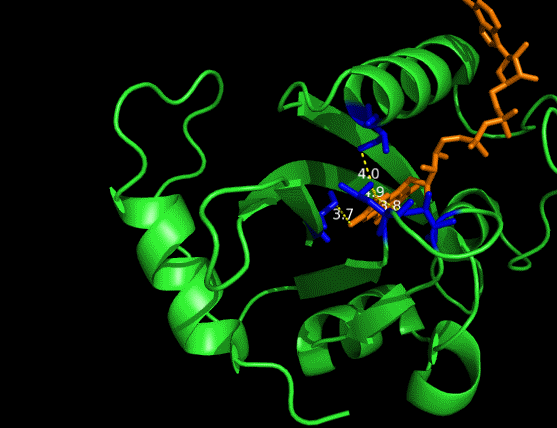
Figure 2. Vancancy in the pocket of VVD[2]
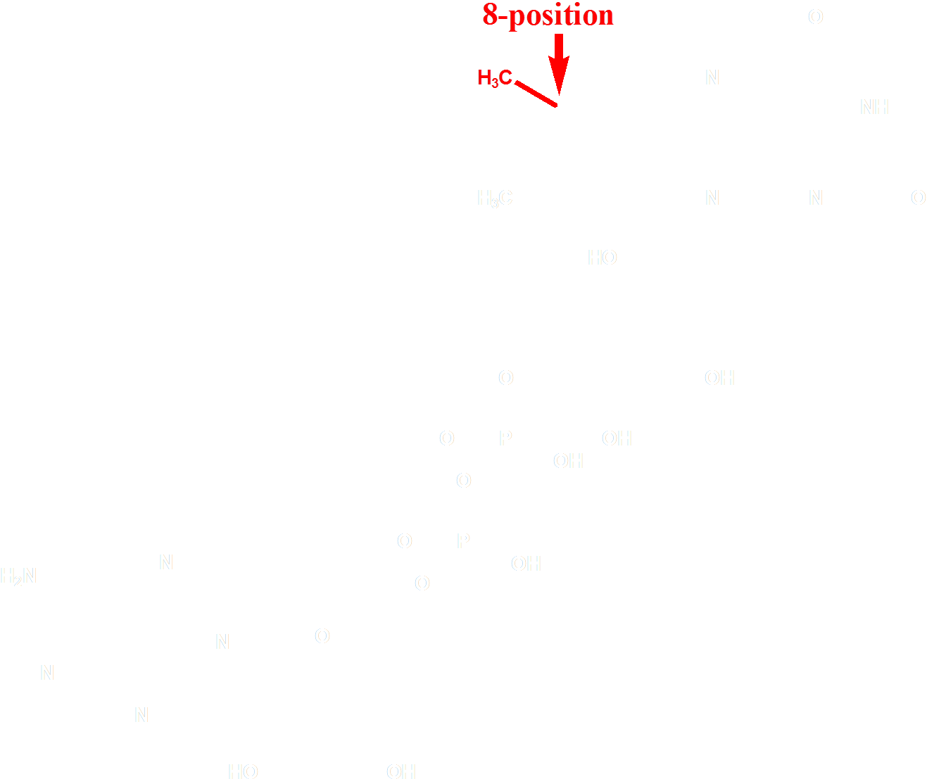
Figure 3. Molecular structure of FAD
Design
The choice of the FAD analogue should have the following features: a red-shifted absorption spectrum and a substitution at 8 position. After selecting from of a pool of over 500 chemicals listed by similarity search on SciFinder, we identified 8-tetrahydropyrroriboflavin (Fig. 4) as a promising candidate for incorporation, which has a red shift of absorption at 510nm.
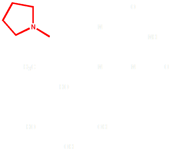
Figure 4 Molecular Structure of 8-tetrahydropyrrole riboflavin
On the other hand, in order to attain bio-orthoganality and to efficiently accommodate the FAD analogue, necessary mutations should be done with the pocket of VVD. Molecular docking results (Fig. 4) indicate that this analogue interacts with the protein at four specific sites(residues marked in blue). These side chains serve as spatial hindrances in that they are within a distance of 3 angstrom (dash lines). Mutations of these residues will hopefully create enough space for the 8-substituted FAD analogue, in this case, a tetrahydropyrrole is at the 8 position.
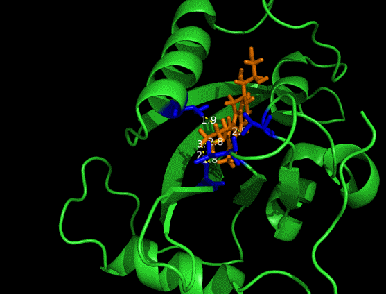
Figure 5 Spatial interactions between FAD analogue and the pocket of VVD[2]
Prospective
A newly developed, dual channel photosensing system based on LexA-VVD will greatly broaden the territory of optogentics. A red-shifted Luminesensor could be used in deep tissue phototherapy, non-invasive light control of neuron and many other fields. What’s more, the concept used in this design could be instructive for future endeavors in developing a spectroscopically different photosensor.
Reference
- 1. Wu Y. et al.(2012). Dynamic Modeling of Human 5-Lipoxygenase−Inhibitor Interactions Helps To Discover Novel Inhibitors. J. Med. Chem., 55: 2597: 605
- 2. Zoltowski, B.D. et al.(2007). Conformational switching in the fungal light sensor Vivid. Science, 316: 1054-1057
- 3. Wang, X., Chen, X., and Yang, Y.(2012). Spatiotemporal control of gene expression by a light-switchable transgene system. Nature Methods, 9:266–269.


