Team:Evry/Tadpole injection1
From 2012.igem.org
Plasmid injection in fertilized Xenopus tropicalis eggs
Plasmids injected:
We injected 2.3 nl of plasmid at 100ng.µl-1 - embryos were stored at 21°C during all the experiment
- pCS2+ GFP-aid: contains the constitutive promoter CMV and the aid sequenced of the aid system fusionned to GFP (Green Fluorescent Protein)(Nishimura et al., 2009). Number of Plasmids injected: ~ 3.78E+7
- pCS2+ citrine: contains the constitutive promoter CMV and the fluorescent protein citrin (yellow). Number of Plasmids injected: ~ 4.38E+7
- pCS2+ mCFP: contains the constitutive promoter CMV and the fluorescent protein mCFP (Cyan Fluorescent Protein). Number of Plasmids injected: ~ 4.37E+7
- pCS2+ GFP-aid
- pCS2+ citrine
- pCS2+ mCFP
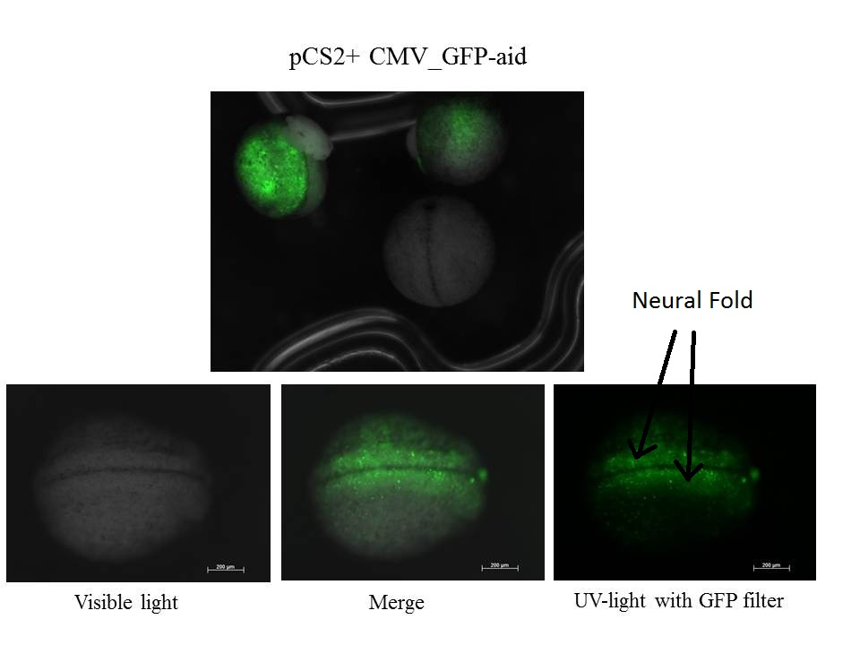
24h after injection
Eggs are near stage 20, neural fold is visible


z-stack of the embryo
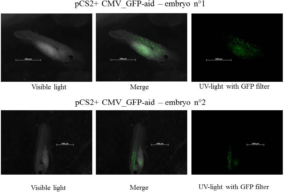
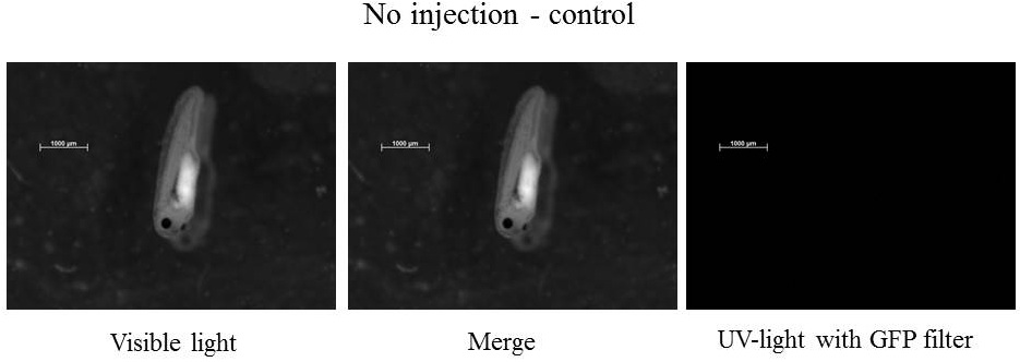
48h after injection
Embryos are at stage 34-38 and move by intermittence


The expression of GFP-aid is localized in different tissue for each tadpole, in spite of the promoter is constitutive (CMV). We can think that the plasmid does not diffuse in the eggs because of the vitellus viscosity.
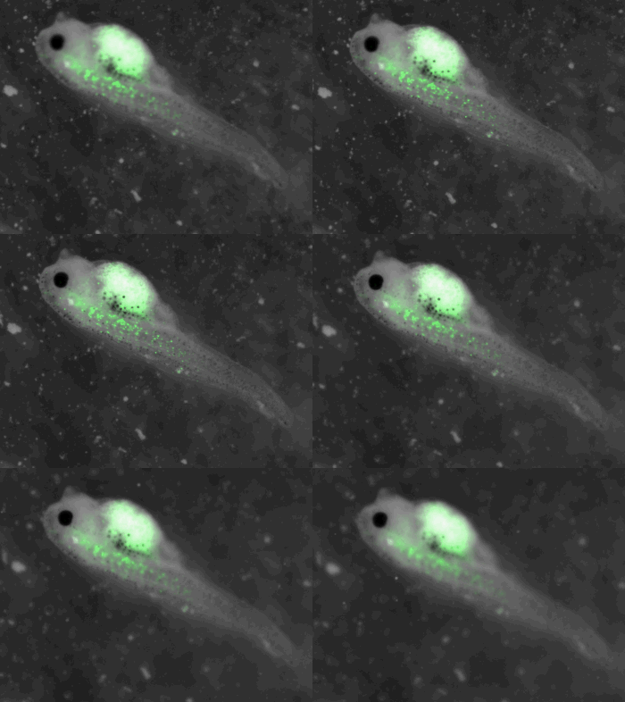
3 days after injection
Embryos are at stage 41-42 and swim


z-stack pictures: pCS2+ CMV_GFP-aid, GFP expression is not in same tissue between tadpoles: for example the tadpole on the left picture bones of the tail produced GFP, on the right picture the GFP expression is localized in the skin. The only part of the tadpole moving is the heart beatting
References:
Nishimura, K. et al., 2009. An auxin-based degron system for the rapid depletion of proteins in nonplant cells. Nature Methods, 6(12), p.917-922.
 "
"