Team:TU-Eindhoven/Notebook/Week10
From 2012.igem.org

Things we did this week
This week, some of us got a guided tour at the building of the Discovery Festival in Eindhoven. The organization is still progressing! Furthermore, everybody is working hard on writing the pieces for the wiki. The [http://www.cursor.tue.nl/nieuwsartikel/artikel/lichtgevende-afbeeldingen-maken-van-gistcellen/ Cursor], the news paper of the Eindhoven University of Technology, published an article on our project. You can find the English verion [http://www.cursor.tue.nl/en/news-article/artikel/lichtgevende-afbeeldingen-maken-van-gistcellen/ here].
Progression in the lab

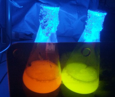
Since we need extra GECO protein for testing, we precultured some E. coli BL21 carrying the GECOs on the pET28a production vector. The precultures are transferred to 100 ml production volumes and induced with IPTG. Because the cooling of the incubator had not been switched on, the temperature was higher than we wanted. It seemed that no protein had been produced in the Erlenmeyer flasks, so we discarded them in the appropriate waste box, waiting for sterilization. A couple of hours later, we discovered that our Erlenmeyer flasks had turned red and green. Therefore we decided to test their fluorescence anyway. Both the R- and the G-GECO are strongly fluorescent in the blue light. We would like to know why the culture was fluorescent after taking it out of the incubator and letting it stand at room temperature for a few hours! Our first guess is that the bacteria have sedimented, started dying of a lack of oxygen at the bottom of the flask and thus releasing their cell contents into the medium. When the released GECOs came into contact with any calcium present in the medium, they got activated. Furthermore, the cell bodies have all sedimented and the medium turned from turbid to clear, allowing fluorescent light to shine colorfully instead of scatter into a blurry white.
We continued working on the BioBricksTM. The colonies that seem to carry the BioBrickTM plasmid are amplified, isolated by miniprep and submitted for sequencing.
How to... describe a model?
We started to describe and explain our model briefly since we have to finish the complete wiki information. Luckily we already had to write a complete bachelor thesis, though describing a complete research is really different from describing your model on just one page of a wiki. Next to it, we still think about applying a sensitivity analysis on our model, but time is running out...
 "
"