Contents |
Transfections


Flowcytometry
Detecting LovTAP
Talk about shifting peak (and why absent in PBS one: dsRed is degraded by formaldehyde)
Characterizing LovTAP
Non-leakage DsRed, where art thou?
Melanopsin Calcium Imaging
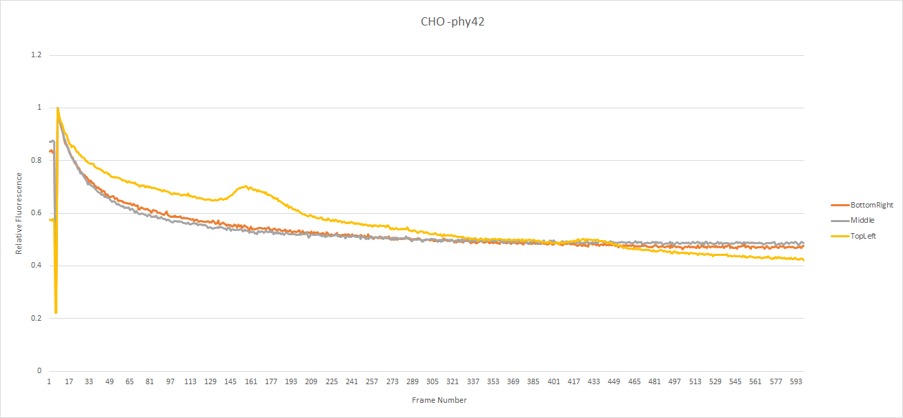
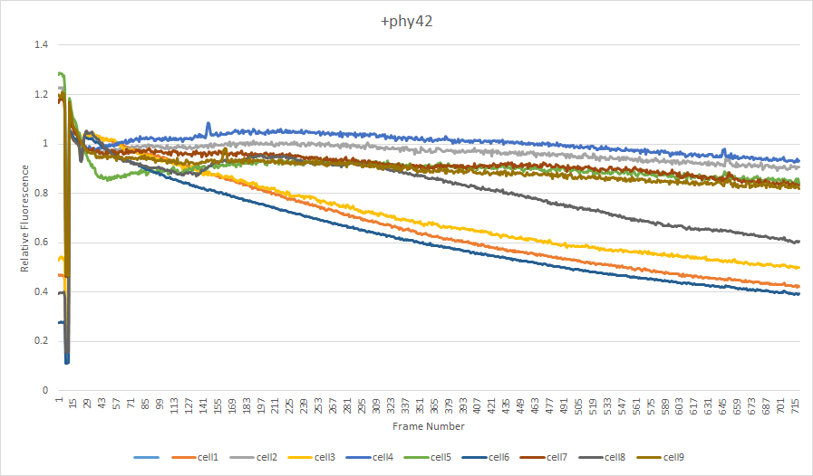
By using timelapse photography and fluorescence microscopy we were able to study the influx of calcium into CHO cells. The cells were stained with a calcium activated dye that fluoresces green, oregon green ( OGB-1/AM ), and exposed to blue light under the microscope.
The CHO seed cells show the expected response to light exposure. The dye is bleached and its intensity gradually decreases along an exponential curve over time. This would seem to indicate the concentration of calcium in the cell remained constant during light exposure and the dye was gradually bleached. The green line seems to go up a bit at certain points, this is due to a cell passing over (out of focus), which increased the local brightness (but, of course, didn't change the real fluorescence).
The CHO cells transfected with PHY42 contain human melanopsin. The cells show a slow linear decrease in dye intensity. This would seem to indicate that calcium is being imported into the cytoplasm while the dye is being bleached.
Part assembly
We planned to clone several parts we used into pSB1C3, which is the standard backbone plasmid in which parts are shipped to the Registry. Four parts were successfully inserted based on colony PCR results followed by successful sequencing.
LovTAP-VP16: BBa_K838000
Successfully cloned 4th of September.
LovTAP readout (RO): BBa_K838001
Successfully cloned on 18th of September
Melanopsin: BBa_K838002 Successfully cloned on 12th of September
 "
"

