Membrane Rudder
Background
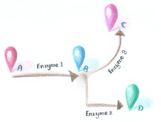
 Fig.1 :Demonstration of branched reactions. Membrane Accelerator employed constitutively dimerizing proteins to assemble enzymes. Imagine if we replace those constitutively dimerizing proteins with signal-induced dimers, which could act as signal sensor, then it is possible to dynamically control the direction of branched reactions. As is described in Figure 1, when signal that induces the dimerization of Enzyme Complex 1 and 2 is present, they would dimerize, making product D more dominant. On the contrary, when signal that induces the dimerization of Enzyme Complex 1 and 3 is present, they would aggregate, so product C is more dominant.
There are indeed many signal induced dimers that commonly exist in nature, which could sense light, chemical, peptide or even RNA signal.
Light Signal
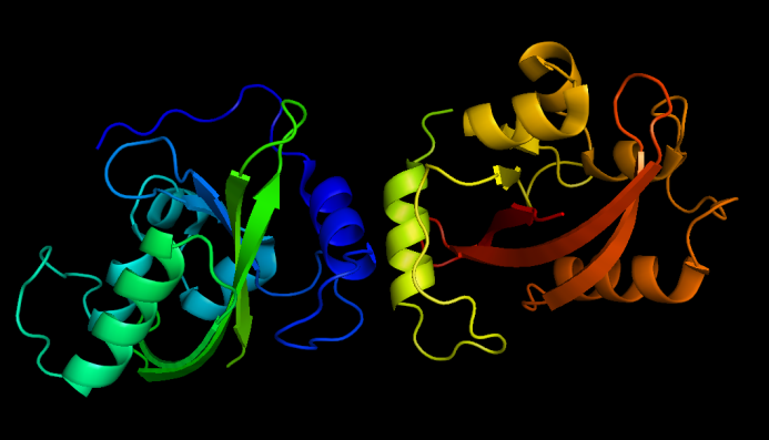
 Fig.2 :Dark state VVD dimers, from PDB ID 2PD7 Light is an intriguing signal to regulate E.coli activity because it is easy to obtain, highly tunable and nontoxic. A metabolite-coupled light-switchable system could be quite fascinating.
Vivid(VVD) protein, a photoreceptor of Neurospora crassa can form dimer in the presence of blue light and disassociate as light is off. Besises, VVD protein belongs to the Per-Arnt-Sim(PAS) protein superfamily.
Compared with wildtype VVD, VVD mutant (C71V and N56K), [http://partsregistry.org/Part:BBa_K771109 Part:BBa_K771109], is harder to dimerize in the dark and easier to dimerize under blue light . So this mutant is ideal as light sensor in Membrane Rudder device. The VVD construction connected to original Membrane Anchor is [http://partsregistry.org/Part:BBa_K771005 Part:BBa_K771005] and [http://partsregistry.org/Part:BBa_K771006 Part:BBa_K771006], for Membrane Anchor 5 and 6.
Whether blue light is present or not could determine which enzymes be assembled to make their final product dominate.
Transcription Signal
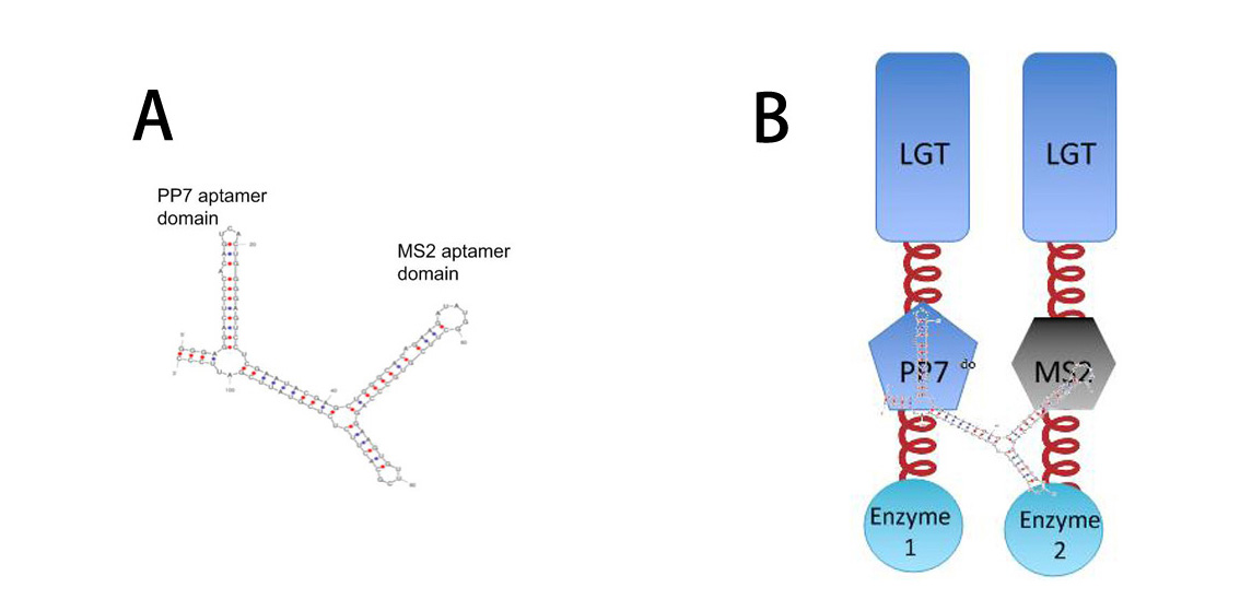
 Fig.10 :A: Sketch of signal RNA molecule (called D0) which consists of PP7 and MS2 aptamer domains. B: Through fusing PP7 and MS2 protein to our membrane device, RNA molecule in Figure A can function as a bridge to connect different proteins, thus decreasing distance between corresponding proteins. So far, Membrane Accelerator and Membrane Rudder is generally about post-translational control over metabolic flux of the host cell. To connect this relatively isolated system to its genetic circuits, we employed RNA signal, which is present in cytoplasm. When certain RNA molecule with dimerization domain is present in cells, its cognate binding proteins can thus dimerize with each other, accelerating certain pathway.
More strikingly, by now, if we want to assemble certain proteins through external signal, we need to find corresponding signal-induced dimer or oligomer. But if we place RNA D0 (with PP7 and MS2 aptamer domains) under control of various promoters regulated by different signals, approaches to induce dimerization would be expanded sharply. Thus, Membrane Rudder could sense much more signals.
Chemical & Peptide Signal
There are many ligand-induced dimers in nature. ErbB family proteins are typically ligand-induced dimers. Estrogen receptor only forms dimer when estrogen is present. If incorporated into our system, it might have significant application prospect. BMP receptors could form heterodimer in the presence of their cognate ligand.
Many other receptors or transport proteins could also form chemical-signal induced dimers.
Recruiting those chemical signal induced dimers could easily broaden the application field of our system.
Summary
What we want to offer is a universal tool which can be connected to different downstream enzymes and upstream signal sensors.
We chose light sensor protein -- VVD to test the feasibility of Membrane Rudder device because of the uniqueness of light signal. Light signal is easily obtained,
Violacein synthetic pathway, a branched enzymatic reaction is recruited to prove Membrane Rudder sensitive to light could work as expected.
Reference
1.Delebecque, C. J., A. B. Lindner, et al. (2011). "Organization of intracellular reactions with rationally designed RNA assemblies." Science 333(6041): 470.
2.Kumar, V. and P. Chambon (1988). "The estrogen receptor binds tightly to its responsive element as a ligand-induced homodimer." Cell 55(1): 145.
3.Lemmon, M. A. (2009). "Ligand-induced ErbB receptor dimerization." Experimental cell research 315(4): 638-648.
4.Terrillon, S. and M. Bouvier (2004). "Roles of G-protein-coupled receptor dimerization." EMBO reports 5(1): 30-34.
5.Wang, X., X. Chen, et al. (2012). "Spatiotemporal control of gene expression by a light-switchable transgene system." Nature Methods.
|