Team:LMU-Munich/Spore Coat Proteins/purification
From 2012.igem.org
Franzi.Duerr (Talk | contribs) |
|||
| Line 39: | Line 39: | ||
{| style="color:black;" cellpadding="0" width="90%" cellspacing="0" border="0" align="center" style="text-align:center;" | {| style="color:black;" cellpadding="0" width="90%" cellspacing="0" border="0" align="center" style="text-align:center;" | ||
|style="width: 70%;background-color: #EBFCE4;" | | |style="width: 70%;background-color: #EBFCE4;" | | ||
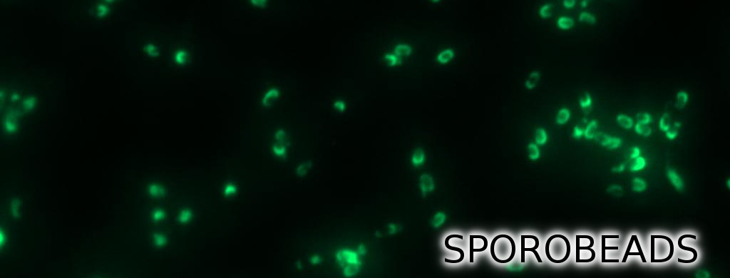
| - | <font color="#000000"; size="2"><p align="justify">Fig. 2: Fluorescence of wild type spores and '''Sporo'''beads after treatment with lysozyme. '''Sporo'''beads still show | + | <font color="#000000"; size="2"><p align="justify">Fig. 2: Fluorescence of wild type spores and '''Sporo'''beads after treatment with lysozyme. '''Sporo'''beads still show undiminished fluorescence activity. </p></font> |
|} | |} | ||
|} | |} | ||
Latest revision as of 13:10, 26 October 2012

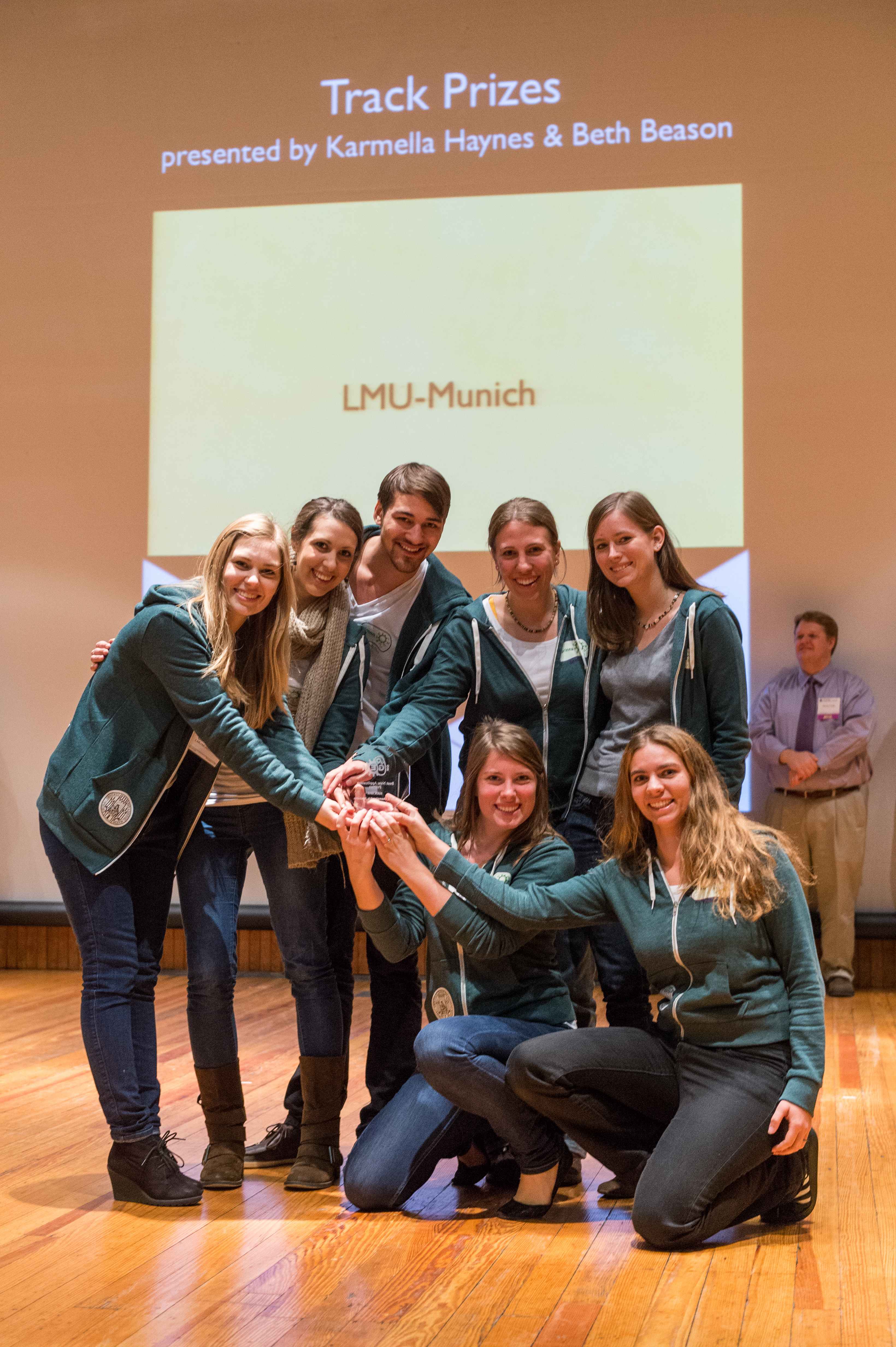
The LMU-Munich team is exuberantly happy about the great success at the World Championship Jamboree in Boston. Our project Beadzillus finished 4th and won the prize for the "Best Wiki" (with Slovenia) and "Best New Application Project".
[ more news ]

Purification Methods
Since there were still some vegetative cells left after 24 hours of growth in Difco Sporulation Medium, we wanted to purify the Sporobeads. We tried three different methods for this approach: treatment with French Press, sonification and lysozyme. By means of the microscopy results we were able to conclude that lysozyme treatment was the only successful method (see data). Additionally, this treatment did not harm the crust fusion proteins as green fluorescence was still detectable afterwards (see data for details).
|
Next, we tested the fluorescence of Sporobeads after lysozyme treatement. This analysis revealed that lysozyme did not harm the gfp-fusion proteins, since fluorescence was not altered (see Fig. 2).
|
 "
"


