 "
"
Team:SDU-Denmark/labwork/Notebook/week3
From 2012.igem.org
| Line 249: | Line 249: | ||
<!-- /// velkomst ////--> | <!-- /// velkomst ////--> | ||
| - | < | + | <h1>Laboratory Notebook</h1> |
| - | + | ||
| - | + | ||
| - | + | ||
<table border="1" bordercolor="#FE1919" style="background-color:#EDEDED" width="100%" cellpadding="3" cellspacing="3"> | <table border="1" bordercolor="#FE1919" style="background-color:#EDEDED" width="100%" cellpadding="3" cellspacing="3"> | ||
| Line 286: | Line 284: | ||
| - | <p> <b> | + | <p> <b>16-07-2012 to 22-07-2012</b> </p> |
<h2>Another digestion, plated SST cultures and Miniprep on FFT </h2> <br/> | <h2>Another digestion, plated SST cultures and Miniprep on FFT </h2> <br/> | ||
<p> | <p> | ||
| Line 295: | Line 293: | ||
</br> | </br> | ||
We also made <a href="https://2012.igem.org/Team:SDU-Denmark/labwork/Protocols/Miniprep">Miniprep</a> on the 10 liquid colonies with FFT, then digested it with EcoRI and PstI. </br> | We also made <a href="https://2012.igem.org/Team:SDU-Denmark/labwork/Protocols/Miniprep">Miniprep</a> on the 10 liquid colonies with FFT, then digested it with EcoRI and PstI. </br> | ||
| - | The digest was | + | The digest was run on a gel and proved results from FFT coloni 3 and 9 which had two bands corrosponding to our vector and gene. </br> |
| - | We measured the concentration on the two tubes using the nano- | + | We measured the concentration on the two tubes using the nano-drop and got the following concentrations: </br> |
<b>Tube#3:</b> 138,2ng/µl </br> | <b>Tube#3:</b> 138,2ng/µl </br> | ||
<b>Tube#9:</b> 71,8ng/µl </br> | <b>Tube#9:</b> 71,8ng/µl </br> | ||
This was enough reason to send them off for sequencing. </br> | This was enough reason to send them off for sequencing. </br> | ||
| - | Seeing as we needed to send of four tubes per sample with 15µl per tube, we didn’t have enough volume in tube#9, so we only prepared four tubes to be send off for sequencing of tube#3, as it had a slightly higher concentration than the 100ng/µL maximum | + | Seeing as we needed to send of four tubes per sample with 15µl per tube, we didn’t have enough volume in tube#9, so we only prepared four tubes to be send off for sequencing of tube#3, as it had a slightly higher concentration than the 100ng/µL maximum. |
</br> | </br> | ||
Furthermore we made 8 new coloni PCR’s from the original FFT amp. agar plate. | Furthermore we made 8 new coloni PCR’s from the original FFT amp. agar plate. | ||
| - | But we only got very small | + | But we only got very small bands on the gel so it wasn't a success. </br> |
For the sequencing, we need four primers, a pJET1.2_for and _rev and then 2 primers that would anneal on the FFT gene. We used the MWG sites primer design tool (http://www.eurofinsdna.com/) to design primers that anneal to the FFT gene. </br> | For the sequencing, we need four primers, a pJET1.2_for and _rev and then 2 primers that would anneal on the FFT gene. We used the MWG sites primer design tool (http://www.eurofinsdna.com/) to design primers that anneal to the FFT gene. </br> | ||
Seeing as the FFT gene is approximately 1800bp long the primers were designed to anneal somewhere between positions 450-500 and 950-1000 both in the forward direction, to be sure that the entire gene is sequenced. </br> | Seeing as the FFT gene is approximately 1800bp long the primers were designed to anneal somewhere between positions 450-500 and 950-1000 both in the forward direction, to be sure that the entire gene is sequenced. </br> | ||
| - | Of the 10 random cultures that were put into liquid LB and left in incubator O.N. on 18/7 only tubes 3 and 9 were viable. We decided to make some more liquid culture of these bacteria and let them incubator at 37°C O.N. </br> | + | Of the 10 random cultures that were put into liquid LB and left in the incubator O.N. on 18/7, only tubes 3 and 9 were viable. We decided to make some more liquid culture of these bacteria and let them incubator at 37°C O.N. </br> |
</br> | </br> | ||
This is to be sure that tomorrow we have enough sample/volume of tube#9 in order to send it off for sequencing. | This is to be sure that tomorrow we have enough sample/volume of tube#9 in order to send it off for sequencing. | ||
| Line 313: | Line 311: | ||
<br/> | <br/> | ||
| - | + | ||
<h2>DNA extraction from O.N. liquid cultures </h2> <br/> | <h2>DNA extraction from O.N. liquid cultures </h2> <br/> | ||
<p> | <p> | ||
| Line 327: | Line 325: | ||
<br/> | <br/> | ||
| - | + | ||
| - | <h2>Gel electrophoresis on extracted | + | <h2>Gel electrophoresis on extracted cultures of FFT and SST </h2> <br/> |
<p> | <p> | ||
We did a cryo “backup” of the liquid cultures from yesterday, containing the FFT and SST genes. | We did a cryo “backup” of the liquid cultures from yesterday, containing the FFT and SST genes. | ||
| Line 335: | Line 333: | ||
The bacteria cultures was lysed and the plasmids was extracted using GeneJET Plasmid Miniprep Kit. | The bacteria cultures was lysed and the plasmids was extracted using GeneJET Plasmid Miniprep Kit. | ||
A Nanodrop analysis was made from our extracted plasmids which should contain our FFT- and SST-gene. | A Nanodrop analysis was made from our extracted plasmids which should contain our FFT- and SST-gene. | ||
| - | A gel was constructed and our plasmids was | + | A gel was constructed and our plasmids was run through at 100V. |
| - | |||
Revision as of 18:46, 25 September 2012


Laboratory Notebook
16-07-2012 to 22-07-2012
Another digestion, plated SST cultures and Miniprep on FFT
The plated SST from yesterday didn’t give any colonies. We tried a different procedure where we use the PCR purification from SST and the pJET plasmid digested with EcoRV in a gel.
We then made a gel purification on both bands at the same time.
The purification was eluted in 30μl elution buffer, and then we used 7μl of the elution with 0,5μl ligase and 2,5μl ligase buffer in order to ligate the gene into the vector.
This was plated on amp-agar plates and incubated O.N.
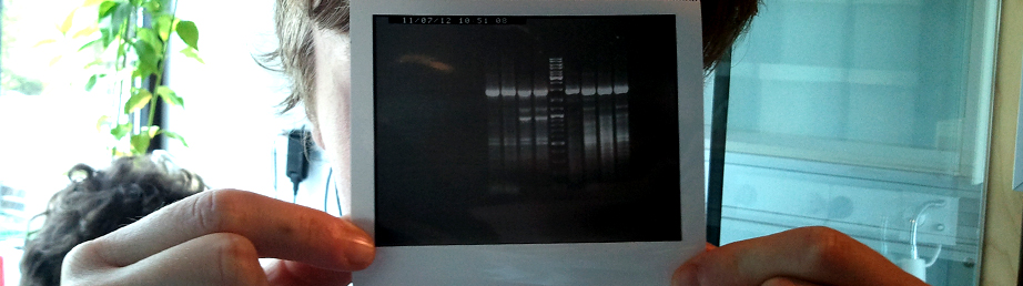
We also made Miniprep on the 10 liquid colonies with FFT, then digested it with EcoRI and PstI.
The digest was run on a gel and proved results from FFT coloni 3 and 9 which had two bands corrosponding to our vector and gene.
We measured the concentration on the two tubes using the nano-drop and got the following concentrations:
Tube#3: 138,2ng/µl
Tube#9: 71,8ng/µl
This was enough reason to send them off for sequencing.
Seeing as we needed to send of four tubes per sample with 15µl per tube, we didn’t have enough volume in tube#9, so we only prepared four tubes to be send off for sequencing of tube#3, as it had a slightly higher concentration than the 100ng/µL maximum.
Furthermore we made 8 new coloni PCR’s from the original FFT amp. agar plate.
But we only got very small bands on the gel so it wasn't a success.
For the sequencing, we need four primers, a pJET1.2_for and _rev and then 2 primers that would anneal on the FFT gene. We used the MWG sites primer design tool (http://www.eurofinsdna.com/) to design primers that anneal to the FFT gene.
Seeing as the FFT gene is approximately 1800bp long the primers were designed to anneal somewhere between positions 450-500 and 950-1000 both in the forward direction, to be sure that the entire gene is sequenced.
Of the 10 random cultures that were put into liquid LB and left in the incubator O.N. on 18/7, only tubes 3 and 9 were viable. We decided to make some more liquid culture of these bacteria and let them incubator at 37°C O.N.
This is to be sure that tomorrow we have enough sample/volume of tube#9 in order to send it off for sequencing.
DNA extraction from O.N. liquid cultures
Overnight cultures incubated at 37°C in Ampicilin containing medium:
- 10 FFT liquid cultures
- 6 SST liquid cultures
The 16 cultures was extracted and a Nanodrop measured the concentrations:
Run 3: 116,1 ng/µl
Run 9: 10,8 ng/µl (discarded, useless for sequencing at this low concentration)
For the preparation of liquid culture for overnight incubation we marked and transferred single-unit colonies from FFT plates(10) and SST plates(6) to 3mL ampicilin containing liquid medium.
Gel electrophoresis on extracted cultures of FFT and SST
We did a cryo “backup” of the liquid cultures from yesterday, containing the FFT and SST genes. The bacteria cultures was lysed and the plasmids was extracted using GeneJET Plasmid Miniprep Kit. A Nanodrop analysis was made from our extracted plasmids which should contain our FFT- and SST-gene. A gel was constructed and our plasmids was run through at 100V. There were no usable results from the gel electrophoresis. For future studies we should try to digest the plasmids with the restriction enzymes for a longer period.