Team:Peking/Project/Overview
From 2012.igem.org
Spring zhq (Talk | contribs) |
Spring zhq (Talk | contribs) |
||
| Line 38: | Line 38: | ||
</div> | </div> | ||
<p> | <p> | ||
| - | + | After primary construction of fusion protein, protein structure inspection and kinetic simulations were conducted to rationally perform optimization. (If you would like to know the details of the <i>Luminesensor</i> design, please see the <a href="/Team:Peking/Project/Luminesensor/Design">Design page</a>.) | |
<br /> <br /> | <br /> <br /> | ||
Due to the consideration of minimal influence on endogenous components, a special mutant of LexA was used to ensure the orthogonality with the endogenous LexA protein. The orthogonal <i>Luminesensor</i> proved to work very well in the common <i>E. coli</i> strain without LexA deletion (data in <a href="/Team:Peking/Project/Luminesensor/Characterization">Characterization</a>). | Due to the consideration of minimal influence on endogenous components, a special mutant of LexA was used to ensure the orthogonality with the endogenous LexA protein. The orthogonal <i>Luminesensor</i> proved to work very well in the common <i>E. coli</i> strain without LexA deletion (data in <a href="/Team:Peking/Project/Luminesensor/Characterization">Characterization</a>). | ||
Revision as of 10:07, 20 September 2012
Achievements
This summer, we have
1. helped 4 other iGEM teams by sharing DNA materials, characterizing their parts and modeling!
2. outlined and detailed a new approach of human pratice called "Sowing Tomorrow Synthetic Biologists"!
3. presented all fresh iGEMers with a collection and praise of historic iGEM projects to share and learn from each other!
4. submitted 11 high quality and well-characterized standard biobricks!
5. improved the ease-of-use of "Lux Brick" that primarily constructed by Cambridge iGEM 2010 and carefully characterized its dynamics of functionality!
6. rationally constructed an ultra-sensitive genetically encoded sensor of luminance – what we call the Luminesensor and comprehensively characterized it. Luminesensor proved to be as sensitive to sense natural light and even bioluminescence!
7. successfully implemented spatiotemporal control of cellular behavior, such as high-resolution 2-D and 3-D bio-printing using dim light, and even utilizing the luminescence of an iPad!
8. successfully implemented cell-cell communication using light for the very first time in synthetic biology!
Therefore, we believe that we deserve a Gold Medal Prize.
1. Luminesensor
-- An Ultrasensitive Light Sensor
Based on comprehensive literature review, Peking iGEM has concluded that an ultra-sensitive light-sensing transcription factor may circumvent serious issues of current optogenetic methods, e.g. cytotoxicy, narrow dynamic range, and dependency on laser and exogenous chromophore.
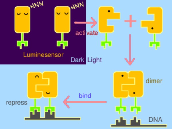
Inspired by the general design principle of genetically encoded sensor in chemical biology, we fused a light-sensing domain, the smallest LOV (light, oxygen, or voltage) protein- Vivid (VVD) from Neurospora, to the DNA binding domain of bacterial LexA, a paradigm of bacterial repressor. When illuminated by blue light (440~480nm), driven by the dimerization of VVD, DNA binding domain of LexA will dimerize and bind to DNA, thus to repress the expression of downstream gene (Figure 1).

Figure 1. The basic design of Luminesensor.
After primary construction of fusion protein, protein structure inspection and kinetic simulations were conducted to rationally perform optimization. (If you would like to know the details of the Luminesensor design, please see the Design page.)
Due to the consideration of minimal influence on endogenous components, a special mutant of LexA was used to ensure the orthogonality with the endogenous LexA protein. The orthogonal Luminesensor proved to work very well in the common E. coli strain without LexA deletion (data in Characterization).
Consistent with the results of kinetic simulation (details in Modeling), mutagenesis based on previous kinetic data of VVD mutation largely improved the property of the Luminesensor, now with an amazing dynamic range reaching 300(data in Characterization).
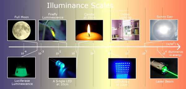
The high sensitivity of the VVD protein is preserved when coupled with the LexA DNA binding domain. Amazingly, the Luminesensor proves to be sensitive to natural light and even bioluminescence (data in Characterization). Such high sensitivity allows the Luminesensor to respond to light signals with broad illuminance scales (Figure 2).

Figure 2. The broad illuminance scales that the Luminesensor could respond to.
Unsatisfied yet with the properties of the Luminesensor, we also wants to red-shift the absorption spectrum of VVD through molecular docking (see Modeling) and mutation of VVD (see Extension), which would largely extend the future application of the Luminesensor, e.g. a multicolor Luminesensor control system.
2. Syn Bio in 2D and 3D
-- An Industrial Application
of Luminesensor
Few optogenetics methods have been applied to industrial use until now. To fully demonstrate the precise spatiotemporal manipulation of light and the bright future of optogenetics, Peking iGEM has deep explored the application of the Luminesensor in 2D and 3D bioprinting methods.
High resolution image could be obtained by 2D bioprinting on the plate mixed with Luminesensor (Figure 3), using easily constructed devices (more on 2D Printing).

Figure 3. High resolution 2D bioprinting.
Even more amazingly, sharp images could be printed using the Retina Display of an Apple iPad (see iPrinting), which serves as the interface between biological systems with electrical devices.
A very promising area of the industrial applications of the Luminesensor is 3D printing, due to the high spatiotemporal specificity of light signals. 3D images could be elegantly printed into the plate mixed with the Luminesensor (see 3D Printing). A new design idea of 3D printer, using holographic technology instead of layer-by-layer printing. This could raise a revolution in the 3D printing world. With such a power tool, 3D printing with biological material has endless potentials in the medical field (crazy ideas on Future).
3. Cell-Cell Light Communication
-- A New Generation of Optogenetics
Cell-cell communication based on quorum sensing systems, e.g. AHL, has been largely utilized by synthetic biologists to construct gene circuits with complex functions, e.g. pattern formation and edge detection. However, the diffusion and saturation of quorum sensing chemicals limit the delivery of communication signals, which in turn requires high spatiotemporal resolution and long range interaction.
The ultrasensitivity of the Luminesensor encouraged our team to explore the possibility of cell-cell light communication (Figure 4). By employing the lux operon from V. fisheri as the light sender and the Luminesensor as the light receiver, we successfully demonstrated, for the first time ever, that light communication between cells could be achieved beyond direct physical contact (see video in Results). Through measuring the difference of the reporter gene expression, it was proved that the light of the lux operon was sufficient to trigger the response of the Luminesensor (data in Results).
A complete light communication system with positive and negative control was constructed by modifying the Light-Off system to Light-On system (details in Design), which enables us to achieve more complex functions based on cell-cell light communication (creative ideas in Future).
4. Phototaxis
-- Cell Motion Controlled by Light
What makes the Luminesensor outstanding is not only its ultra-sensitivity, but also the particularly high dynamic range (data on Characterization). Such switch-like property makes the Luminesensor a very reliable and beneficial module in synthetic biology. Similar to the light-controlled animal behavior in neuroscience, by coupling the Luminesensor with endogenous systems, it is possible to integrate the light signal with the native response to control biological behavior on a higher level, e.g. cell motion.
The 2012 Peking iGEM has successfully built "Phototatic" bacteria by programming the chemotaxis system in E. coli through light with the Luminesensor (Figure 5). By controlling the expression level of the cheZ protein with light, the tumbling frequency is coupled to the intensity of light signals (details in Design). On the border of the light and dark fields on the plate, the motility difference of the cells in a single colony on the two sides is sufficient to form an uneven colony (see Demonstration). The light-controlled cell motion that we have achieved has a very promising environmental and medical application for the future, e.g. the delivery of drugs to target concerns.
 "
"


