Team:Georgia Tech/sandbox/Project/
From 2012.igem.org
Elsherbini (Talk | contribs) |
Elsherbini (Talk | contribs) |
||
| Line 35: | Line 35: | ||
==Results== | ==Results== | ||
| - | |||
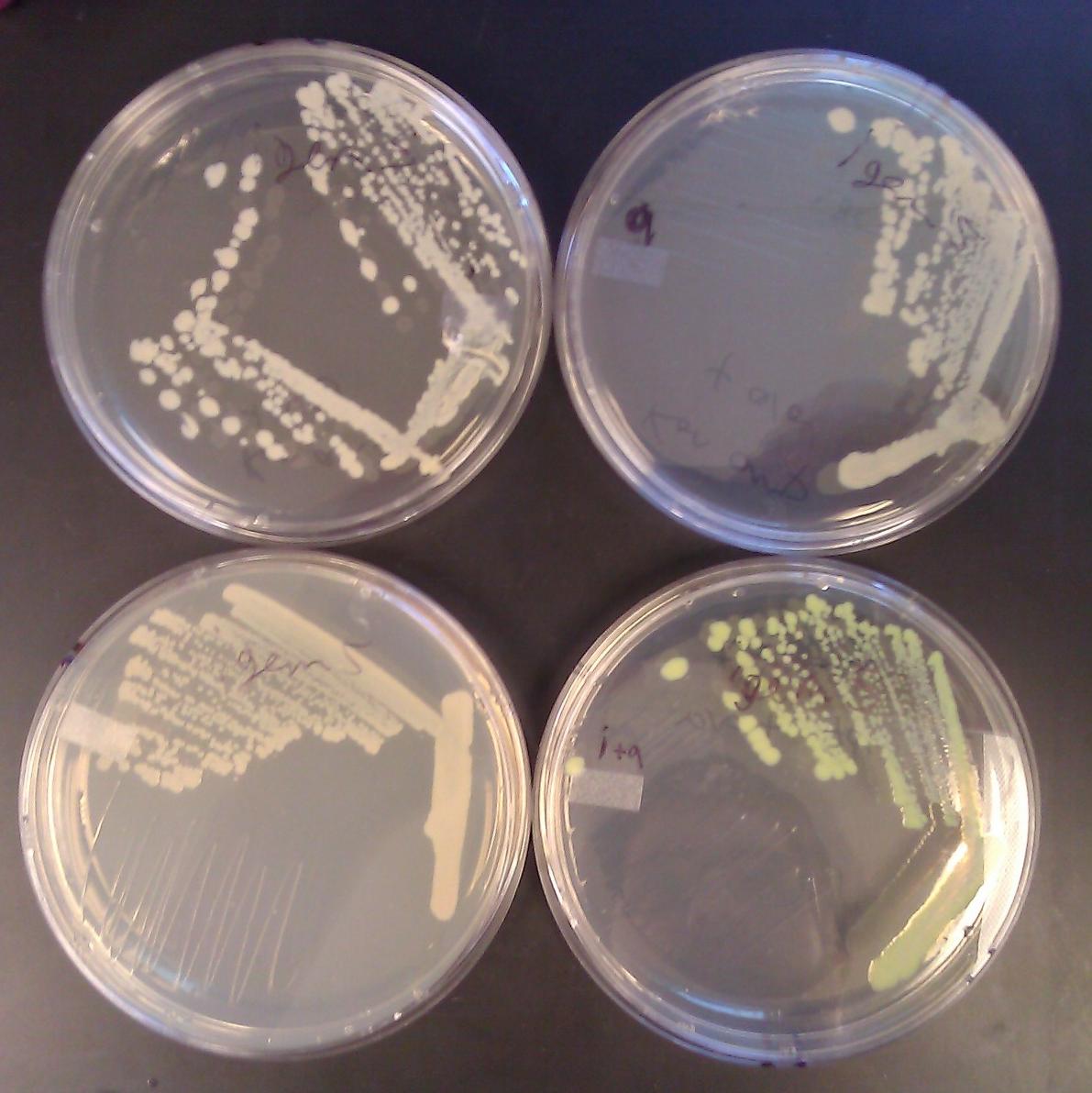
[[File:GT_leucine_zipper_GFP_plates.JPG|350px|thumb|right|Leucine zipper controls glow only upon addition of both Arabinose and IPTG]] | [[File:GT_leucine_zipper_GFP_plates.JPG|350px|thumb|right|Leucine zipper controls glow only upon addition of both Arabinose and IPTG]] | ||
| + | |||
| + | After co-transforming pET11a-z-NGFP and pMRBAD-z-CGFP into BL21(DE3) cells, we plated on media containing IPTG (0.1mM) and arabinose (0.2% w/v). We incubated at 30° C for 10 hours and left the plates on the bench top for 24 hours. We observed accumulation of GFP only in colonies plated with both IPTG and arabinose. | ||
| + | |||
<groupparts>iGEM012 Georgia_Tech</groupparts> | <groupparts>iGEM012 Georgia_Tech</groupparts> | ||
Revision as of 23:02, 2 October 2012
Contents |
Abstract
An intragenic complementation approach to engineer a faster fluorescence biosensor
Our goal is to engineer a novel biosensor with a faster readout than is currently available. Many bacteria produce, secrete, and respond to chemicals called autoinducers to monitor population density and to synchronize gene expression, a process called quorum sensing. In quorum sensing based biosensors, detection of autoinducer activates transcription of a reporter gene, which must then be translated and accumulate to detectable levels, which can take two to four hours. In our system, we will use TraR, a protein used in the quorum sensing response of Agrobacterium tumefaciens, which dimerizes only in the presence of its autoinducer. We have successfully fused traR to sequence for two separate complementary fragments of GFP. Upon addition of autoinducer, we predict that already accumulated TraR-GFP fragment monomers will dimerize, allowing the GFP fragments to interact and fluoresce. This new approach may drastically reduce the time necessary for future biosensors to produce detectable output.
Project Overview
Motivation
Design Considerations
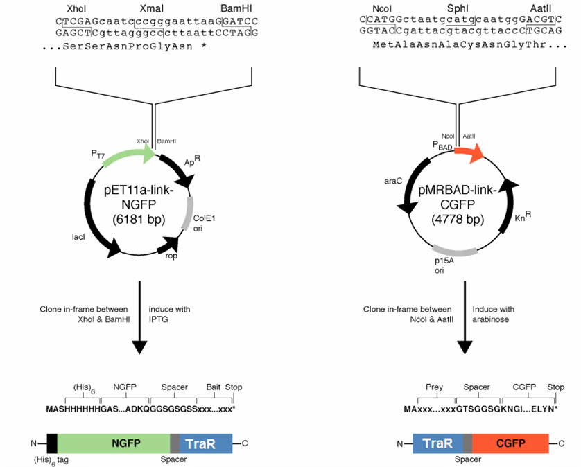
We decided to first make the fusions in the plasmids provided by Dr. Regan from Yale. By choosing this method, we were able to use the provided controls in testing our part.
The designed fusion into pET11a was compatible with the TraR sequence, but the pMRBAD fusion was not. The pMRBAD fusion was designed to use NcoI and AatII, but TraR had two AatII sites in the coding sequence. We instead cloned TraR in-between the NcoI and Sph1 sites. This added two amino acids to the linker, one of which was a cysteine. It is unclear whether these additional amino acids will disrupt the ability for TraR and the CGFP fragment to fold independently from one another.
The junction between traR and the C-terminal GFP fragment cloned into the SphI site:
                   SphI
   R   K   L   I   A   C   N   G   T   S   G
   cgg aaa ctc atc gca tgc aat ggg acg tcg ggt
       <-- traR--> <-- linker -->
Results
After co-transforming pET11a-z-NGFP and pMRBAD-z-CGFP into BL21(DE3) cells, we plated on media containing IPTG (0.1mM) and arabinose (0.2% w/v). We incubated at 30° C for 10 hours and left the plates on the bench top for 24 hours. We observed accumulation of GFP only in colonies plated with both IPTG and arabinose.
<groupparts>iGEM012 Georgia_Tech</groupparts>
 "
"


