Team:ETH Zurich/Decoder/Results
From 2012.igem.org
| (90 intermediate revisions not shown) | |||
| Line 8: | Line 8: | ||
| - | === | + | === Pre-decoder === |
| + | [[File: ethzurich_Decoderpart1_pseva.png|frameless|400px|right]] | ||
In a first step two promoters were cloned together. | In a first step two promoters were cloned together. | ||
| - | The promoter controling mCherry expression is repressible by TetR and cI while the promoter controling | + | The promoter controling mCherry expression is repressible by TetR and cI while the promoter controling eCFP expression is repressible by LacI and cI. To verify the expectations we used the pSEVA-LacI-tetR to test our construct for 4 distinct expression conditions. |
| - | |||
| - | |||
| - | |||
| - | |||
| - | |||
| - | |||
| - | |||
| + | [[File: Experimentsetuppart1ethz2012.png|frameless|300px|right]] | ||
| - | |||
| - | + | TOP10 cells cotransformed with the pSEVA-LacI-tetR and the pre-decoder plasmid were grown in 250 mL shaking flask in LB containing Ampicillin and Kanamycin. After a certain time, each pre-culture was aliquoted into 4 shaking flasks containing: | |
| - | |||
| - | |||
| + | * 1. No inducer: LacI as well as TetR (due to the leakiness of the tac promoter) are present and bind to the Lac & Tet operator regions. No output. | ||
| + | * 2. IPTG: LacI cannot repress the tac promoter anymore, TetR is expressed. TetR represses the expression of mCherry. On the other hand the promoter controling eCFP is active. Blue output. | ||
| - | + | * 3. aTc: LacI is expressed, TetR is not there, mCherry is expressed. eCFP cannot be produced as LacI binds to the LacO operator region. Red output. | |
| + | * 4. IPTG & aTc: mCherry as well as eCFP are expressed. Red and blue output. | ||
| - | + | ||
| - | | [[File: | + | |
| - | | [[File: | + | |
| + | === FACS=== | ||
| + | |||
| + | Single cell analysis revealed that the repression by LacI and TetR occurs and that the promoters work in absence of the repressors. | ||
| + | |||
| + | <div class="eth_imagetable"> | ||
| + | {| | ||
| + | |[[File: 2012ethz_part1decGrowth_curve.png|frameless|300px|right|thumb|Growth curve of the cotransformed TOP10 cells. All of them grew well and similarly fast. The time after induction was measured after aliquotation of the precultures into the 4 differently treated flasks.]] | ||
| + | |[[File:New-Pre-decoder-ohne.png|frameless|300px|right|thumb|The peaks of the four conditions show the expected properties, which is also visible in the scatterplot (over 90% of each population shown). Induction with IPTG leads to eCFP expression, while mCherry is not expressed. Addition of aTc leads to mCherry expression, while no eCFP is produced. IPTG together and aTc lead to expression of both XFPs. No induction does not lead to any XFP expression. The graphs show the data set obtained 4.5 hours after induction.]] | ||
| + | |- | ||
|} | |} | ||
| + | </div> | ||
| + | This is a proof that the both promoters are working and that the operator sites can indeed be repressed by LacI and TetR. With that knowledge we could start assembling the whole decoder and check if the promoters can also be repressed by cI. | ||
| + | |||
| + | |||
| + | == Full decoder == | ||
| + | |||
| + | |||
| + | The following construct was cloned to the first part of the decoder. By that, the third repressor cI is introduced to the system. If the promoter is turned on, cI is expressed and represses the other two hybrid promoters. To test the decoder we used the same procedure as mentioned above. The four conditions are then the following ones: | ||
| + | |||
| + | * 1. No inducer: LacI as well as TetR (due to the leakiness of the tac promoter) are present and bind to the Lac & Tet operator regions. All the promoters are turned off. No output. | ||
| + | |||
| + | * 2. IPTG: LacI cannot repress the tac promoter anymore, TetR is expressed. TetR represses the expression of mCherry, cI and YFP. On the other hand the promoter controling eCFP is active. Blue output. | ||
| + | |||
| + | * 3. aTc: LacI is expressed, TetR is not there, mCherry is expressed. eCFP, cI and YFP cannot be produced as LacI binds to the LacO operator region. Red output. | ||
| + | |||
| + | * 4. IPTG & aTc: cI and YFP are expressed. cI represses mCherry and eCFP. Yellow output. | ||
| + | |||
| + | <div class="eth_imagetable"> | ||
| + | {| | ||
| + | |[[File: ethz2012Tetlacciyfp1.png|thumb|400px|center]] | ||
| + | |[[File: Decoder_wiki_ethz2012.png|thumb|240px|right|Hybrid promoters combined to the decoder. ]] | ||
| + | |} | ||
| + | </div> | ||
| + | |||
| + | |||
| + | |||
| + | A first glimpse at the plates revealed already that the hybrid promoters are repressible by cI. While colonies containing only the hybrid promoters controling mCherry and eCFP were shining mainly red, the ones containing the whole decoder were hardly red. | ||
| + | |||
| + | <div class="eth_imagetable"> | ||
| + | {| | ||
| + | |[[File: 2012ethDecoderall.png|frameless|250px|left|thumb| Top: decoder part1; Left: P(TetO,LacO) controling cI and YFP; Right:Final decoder.]] | ||
| + | |[[File: Decoder_gel_2012ethz.png|frameless|300px|right|thumb|Left to right: Ladder, 3 screened colonies containing the decoder plasmid. ]] | ||
| + | |} | ||
| + | </div> | ||
| - | [[File:Proof-of-principle.jpg|frameless|700px|center|thumb| | + | [[File:Proof-of-principle.jpg|frameless|700px|center|thumb|Boolean logic for testing of Decoder. ]] |
{{:Team:ETH_Zurich/Templates/Footer}} | {{:Team:ETH_Zurich/Templates/Footer}} | ||
Latest revision as of 02:27, 27 October 2012
Contents |
Hybrid promoters
Pre-decoder
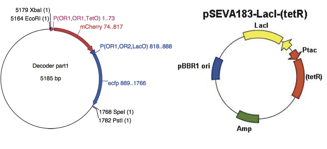
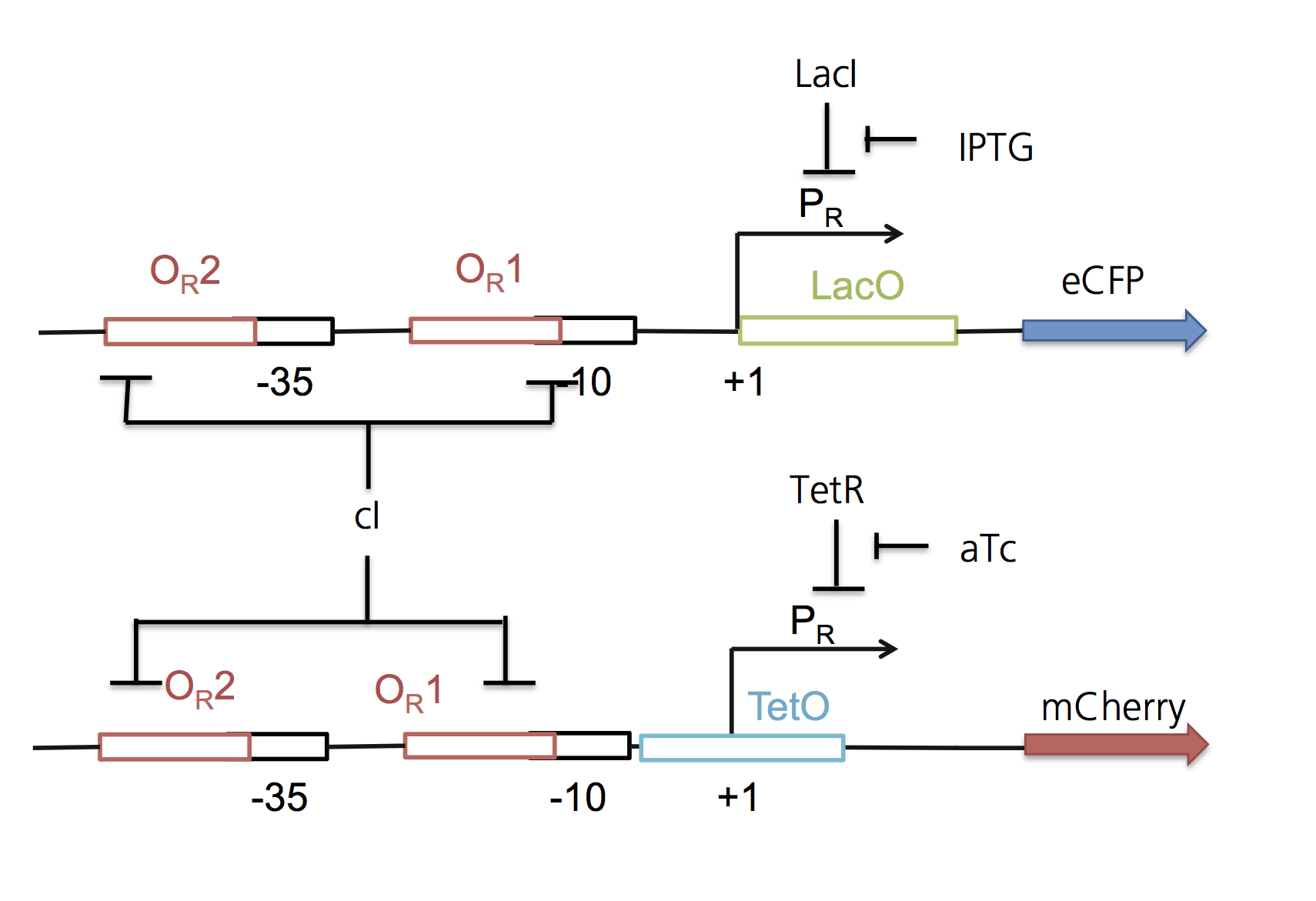
In a first step two promoters were cloned together. The promoter controling mCherry expression is repressible by TetR and cI while the promoter controling eCFP expression is repressible by LacI and cI. To verify the expectations we used the pSEVA-LacI-tetR to test our construct for 4 distinct expression conditions.
TOP10 cells cotransformed with the pSEVA-LacI-tetR and the pre-decoder plasmid were grown in 250 mL shaking flask in LB containing Ampicillin and Kanamycin. After a certain time, each pre-culture was aliquoted into 4 shaking flasks containing:
- 1. No inducer: LacI as well as TetR (due to the leakiness of the tac promoter) are present and bind to the Lac & Tet operator regions. No output.
- 2. IPTG: LacI cannot repress the tac promoter anymore, TetR is expressed. TetR represses the expression of mCherry. On the other hand the promoter controling eCFP is active. Blue output.
- 3. aTc: LacI is expressed, TetR is not there, mCherry is expressed. eCFP cannot be produced as LacI binds to the LacO operator region. Red output.
- 4. IPTG & aTc: mCherry as well as eCFP are expressed. Red and blue output.
FACS
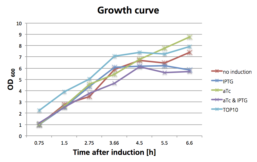
Single cell analysis revealed that the repression by LacI and TetR occurs and that the promoters work in absence of the repressors.
This is a proof that the both promoters are working and that the operator sites can indeed be repressed by LacI and TetR. With that knowledge we could start assembling the whole decoder and check if the promoters can also be repressed by cI.
Full decoder
The following construct was cloned to the first part of the decoder. By that, the third repressor cI is introduced to the system. If the promoter is turned on, cI is expressed and represses the other two hybrid promoters. To test the decoder we used the same procedure as mentioned above. The four conditions are then the following ones:
- 1. No inducer: LacI as well as TetR (due to the leakiness of the tac promoter) are present and bind to the Lac & Tet operator regions. All the promoters are turned off. No output.
- 2. IPTG: LacI cannot repress the tac promoter anymore, TetR is expressed. TetR represses the expression of mCherry, cI and YFP. On the other hand the promoter controling eCFP is active. Blue output.
- 3. aTc: LacI is expressed, TetR is not there, mCherry is expressed. eCFP, cI and YFP cannot be produced as LacI binds to the LacO operator region. Red output.
- 4. IPTG & aTc: cI and YFP are expressed. cI represses mCherry and eCFP. Yellow output.
A first glimpse at the plates revealed already that the hybrid promoters are repressible by cI. While colonies containing only the hybrid promoters controling mCherry and eCFP were shining mainly red, the ones containing the whole decoder were hardly red.
References
- Brown, B. a, Headland, L. R., & Jenkins, G. I. (2009). UV-B action spectrum for UVR8-mediated HY5 transcript accumulation in Arabidopsis. Photochemistry and photobiology, 85(5), 1147–55.
- Christie, J. M., Salomon, M., Nozue, K., Wada, M., & Briggs, W. R. (1999): LOV (light, oxygen, or voltage) domains of the blue-light photoreceptor phototropin (nph1): binding sites for the chromophore flavin mononucleotide. Proceedings of the National Academy of Sciences of the United States of America, 96(15), 8779–83.
- Christie, J. M., Arvai, A. S., Baxter, K. J., Heilmann, M., Pratt, A. J., O’Hara, A., Kelly, S. M., et al. (2012). Plant UVR8 photoreceptor senses UV-B by tryptophan-mediated disruption of cross-dimer salt bridges. Science (New York, N.Y.), 335(6075), 1492–6.
- Cloix, C., & Jenkins, G. I. (2008). Interaction of the Arabidopsis UV-B-specific signaling component UVR8 with chromatin. Molecular plant, 1(1), 118–28.
- Cox, R. S., Surette, M. G., & Elowitz, M. B. (2007). Programming gene expression with combinatorial promoters. Molecular systems biology, 3(145), 145. doi:10.1038/msb4100187
- Drepper, T., Eggert, T., Circolone, F., Heck, A., Krauss, U., Guterl, J.-K., Wendorff, M., et al. (2007). Reporter proteins for in vivo fluorescence without oxygen. Nature biotechnology, 25(4), 443–5
- Drepper, T., Krauss, U., & Berstenhorst, S. M. zu. (2011). Lights on and action! Controlling microbial gene expression by light. Applied microbiology, 23–40.
- EuropeanCommission (2006). SCIENTIFIC COMMITTEE ON CONSUMER PRODUCTS SCCP Opinion on Biological effects of ultraviolet radiation relevant to health with particular reference to sunbeds for cosmetic purposes.
- Elvidge, C. D., Keith, D. M., Tuttle, B. T., & Baugh, K. E. (2010). Spectral identification of lighting type and character. Sensors (Basel, Switzerland), 10(4), 3961–88.
- GarciaOjalvo, J., Elowitz, M. B., & Strogatz, S. H. (2004). Modeling a synthetic multicellular clock: repressilators coupled by quorum sensing. Proceedings of the National Academy of Sciences of the United States of America, 101(30), 10955–60.
- Gao Q, Garcia-Pichel F. (2011). Microbial ultraviolet sunscreens. Nat Rev Microbiol. 9(11):791-802.
- Goosen N, Moolenaar GF. (2008) Repair of UV damage in bacteria. DNA Repair (Amst).7(3):353-79.
- Heijde, M., & Ulm, R. (2012). UV-B photoreceptor-mediated signalling in plants. Trends in plant science, 17(4), 230–7.
- Hirose, Y., Narikawa, R., Katayama, M., & Ikeuchi, M. (2010). Cyanobacteriochrome CcaS regulates phycoerythrin accumulation in Nostoc punctiforme, a group II chromatic adapter. Proceedings of the National Academy of Sciences of the United States of America, 107(19), 8854–9.
- Hirose, Y., Shimada, T., Narikawa, R., Katayama, M., & Ikeuchi, M. (2008). Cyanobacteriochrome CcaS is the green light receptor that induces the expression of phycobilisome linker protein. Proceedings of the National Academy of Sciences of the United States of America, 105(28), 9528–33.
- Kast, Asif-Ullah & Hilvert (1996) Tetrahedron Lett. 37, 2691 - 2694., Kast, Asif-Ullah, Jiang & Hilvert (1996) Proc. Natl. Acad. Sci. USA 93, 5043 - 5048
- Kiefer, J., Ebel, N., Schlücker, E., & Leipertz, A. (2010). Characterization of Escherichia coli suspensions using UV/Vis/NIR absorption spectroscopy. Analytical Methods, 9660. doi:10.1039/b9ay00185a
- Kinkhabwala, A., & Guet, C. C. (2008). Uncovering cis regulatory codes using synthetic promoter shuffling. PloS one, 3(4), e2030.
- Krebs in Deutschland 2005/2006. Häufigkeiten und Trends. 7. Auflage, 2010, Robert Koch-Institut (Hrsg) und die Gesellschaft der epidemiologischen Krebsregister in Deutschland e. V. (Hrsg). Berlin.
- Lamparter, T., Michael, N., Mittmann, F., & Esteban, B. (2002). Phytochrome from Agrobacterium tumefaciens has unusual spectral properties and reveals an N-terminal chromophore attachment site. Proceedings of the National Academy of Sciences of the United States of America, 99(18), 11628–33.
- Levskaya, A. et al (2005). Engineering Escherichia coli to see light. Nature, 438(7067), 442.
- Mancinelli, A. (1986). Comparison of spectral properties of phytochromes from different preparations. Plant physiology, 82(4), 956–61.
- Nakasone, Y., Ono, T., Ishii, A., Masuda, S., & Terazima, M. (2007). Transient dimerization and conformational change of a BLUF protein: YcgF. Journal of the American Chemical Society, 129(22), 7028–35.
- Orth, P., & Schnappinger, D. (2000). Structural basis of gene regulation by the tetracycline inducible Tet repressor-operator system. Nature structural biology, 215–219.
- Parkin, D.M., et al., Global cancer statistics, 2002. CA: a cancer journal for clinicians, 2005. 55(2): p. 74-108.
- Rajagopal, S., Key, J. M., Purcell, E. B., Boerema, D. J., & Moffat, K. (2004). Purification and initial characterization of a putative blue light-regulated phosphodiesterase from Escherichia coli. Photochemistry and photobiology, 80(3), 542–7.
- Rizzini, L., Favory, J.-J., Cloix, C., Faggionato, D., O’Hara, A., Kaiserli, E., Baumeister, R., et al. (2011). Perception of UV-B by the Arabidopsis UVR8 protein. Science (New York, N.Y.), 332(6025), 103–6.
- Roux, B., & Walsh, C. T. (1992). p-aminobenzoate synthesis in Escherichia coli: kinetic and mechanistic characterization of the amidotransferase PabA. Biochemistry, 31(30), 6904–10.
- Strickland, D. (2008). Light-activated DNA binding in a designed allosteric protein. Proceedings of the National Academy of Sciences of the United States of America, 105(31), 10709–10714.
- Sinha RP, Häder DP. UV-induced DNA damage and repair: a review. Photochem Photobiol Sci. (2002). 1(4):225-36
- Sambandan DR, Ratner D. (2011). Sunscreens: an overview and update. J Am Acad Dermatol. 2011 Apr;64(4):748-58.
- Tabor, J. J., Levskaya, A., & Voigt, C. A. (2011). Multichromatic Control of Gene Expression in Escherichia coli. Journal of Molecular Biology, 405(2), 315–324.
- Thibodeaux, G., & Cowmeadow, R. (2009). A tetracycline repressor-based mammalian two-hybrid system to detect protein–protein interactions in vivo. Analytical biochemistry, 386(1), 129–131.
- Tschowri, N., & Busse, S. (2009). The BLUF-EAL protein YcgF acts as a direct anti-repressor in a blue-light response of Escherichia coli. Genes & development, 522–534.
- Tschowri, N., Lindenberg, S., & Hengge, R. (2012). Molecular function and potential evolution of the biofilm-modulating blue light-signalling pathway of Escherichia coli. Molecular microbiology.
- Tyagi, A. (2009). Photodynamics of a flavin based blue-light regulated phosphodiesterase protein and its photoreceptor BLUF domain.
- Vainio, H. & Bianchini, F. (2001). IARC Handbooks of Cancer Prevention: Volume 5: Sunscreens. Oxford University Press, USA
- Quinlivan, Eoin P & Roje, Sanja & Basset, Gilles & Shachar-Hill, Yair & Gregory, Jesse F & Hanson, Andrew D. (2003). The folate precursor p-aminobenzoate is reversibly converted to its glucose ester in the plant cytosol. The Journal of biological chemistry, 278.
- van Thor, J. J., Borucki, B., Crielaard, W., Otto, H., Lamparter, T., Hughes, J., Hellingwerf, K. J., et al. (2001). Light-induced proton release and proton uptake reactions in the cyanobacterial phytochrome Cph1. Biochemistry, 40(38), 11460–71.
- Wegkamp A, van Oorschot W, de Vos WM, Smid EJ. (2007 )Characterization of the role of para-aminobenzoic acid biosynthesis in folate production by Lactococcus lactis. Appl Environ Microbiol. Apr;73(8):2673-81.
 "
"
