Team:LMU-Munich/Spore Coat ProteinsTour/Background
From 2012.igem.org
(Created page with "<!-- Include the next line at the beginning of every page --> {{:Team:LMU-Munich/Templates/Page Header|File:Team-LMU_eppis.resized.jpg|3}} [[File:SporeCoat.png|100px|right|link=...") |
|||
| (7 intermediate revisions not shown) | |||
| Line 1: | Line 1: | ||
<!-- Include the next line at the beginning of every page --> | <!-- Include the next line at the beginning of every page --> | ||
{{:Team:LMU-Munich/Templates/Page Header|File:Team-LMU_eppis.resized.jpg|3}} | {{:Team:LMU-Munich/Templates/Page Header|File:Team-LMU_eppis.resized.jpg|3}} | ||
| + | [[File:LMU Glow Spore2 cutII.jpg|620px|link=]] | ||
| - | [[File:SporeCoat.png|100px|right|link= | + | [[File:SporeCoat.png|100px|right|link=]] |
| - | ===Scientific Background=== | + | ===Scientific Background of our '''Sporo'''beads=== |
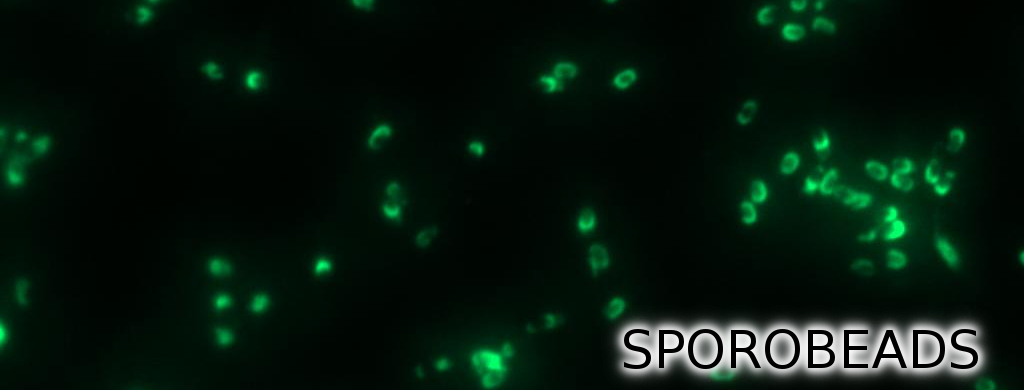
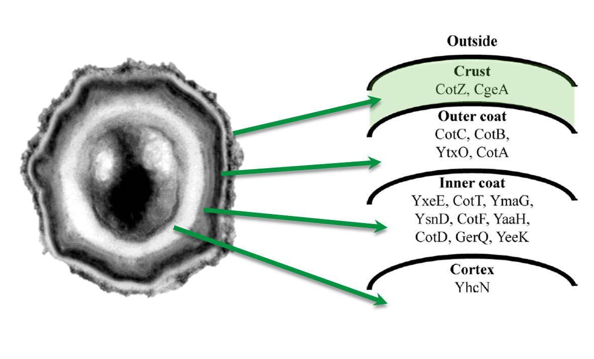
| - | <p align="justify">'''Sporo'''beads are ''Bacillus subtilis'' spores | + | <p align="justify">'''Sporo'''beads are ''Bacillus subtilis'' spores modified so that they can be used as a platform for individual protein display. The aim of this module is to create these spores that display fusion proteins on their surface. There are several different proteins forming the spore coat layers of ''B. subtilis'' spores (Fig. 1). The outermost layer, the so-called spore crust, is composed of two proteins, CotZ and CgeA ([http://www.ncbi.nlm.nih.gov/pubmed?term=imamura%20et%20al.%202011%20spore%20crust Imamura et al., 2011]). This is why we used these two crust proteins to create functional fusion proteins to be expressed on our '''Sporo'''beads.</p> |
| Line 16: | Line 17: | ||
| style="width: 70%;background-color: #EBFCE4;" | | | style="width: 70%;background-color: #EBFCE4;" | | ||
{| | {| | ||
| - | |[[File:Imamura, 2011 & McKenney, 2010.png|Protein distribution in spore coat of ''Bacillus subtilis''|500px|center]] | + | |[[File:Imamura, 2011 & McKenney, 2010.png|Protein distribution in spore coat of ''Bacillus subtilis''|500px|center|link=]] |
|- | |- | ||
| style="width: 70%;background-color: #EBFCE4;" | | | style="width: 70%;background-color: #EBFCE4;" | | ||
| Line 31: | Line 32: | ||
=====Crust Protein Genes Organization===== | =====Crust Protein Genes Organization===== | ||
| - | <p align="justify">The gene ''cgeA'' is located in the ''cgeABCDE'' cluster and is expressed from its own promoter P<sub>''cgeA''</sub>. The cluster ''cotVWXYZ'' contains the gene ''cotZ'' which is cotranscribed with ''cotY'' and expressed from the promoter P<sub>''cotYZ''</sub>. Another promoter of this cluster, P<sub>''cotV''</sub>, is responsible for the transcription of the other three genes (Fig. 2). Those three promoters were evaluated with the ''lux'' reporter genes ( | + | <p align="justify">The gene ''cgeA'' is located in the ''cgeABCDE'' cluster and is expressed from its own promoter P<sub>''cgeA''</sub>. The cluster ''cotVWXYZ'' contains the gene ''cotZ'' which is cotranscribed with ''cotY'' and expressed from the promoter P<sub>''cotYZ''</sub>. Another promoter of this cluster, P<sub>''cotV''</sub>, is responsible for the transcription of the other three genes (Fig. 2). Those three promoters were evaluated with the ''lux'' reporter genes (pSB<sub>''Bs''</sub>3C-''lux''ABCDE) to analyze their strength and the time point of their activation (visit later DATA page). Based on this data, two of the three promoters could be used for expression of spore crust fusion proteins. |
| + | </p> | ||
| Line 37: | Line 39: | ||
| style="width: 100%;background-color: #EBFCE4;" | | | style="width: 100%;background-color: #EBFCE4;" | | ||
{| | {| | ||
| - | |[[File:Operons.png|610px|center]] | + | |[[File:Operons.png|610px|center|link=]] |
|- | |- | ||
| style="width: 70%;background-color: #EBFCE4;" | | | style="width: 70%;background-color: #EBFCE4;" | | ||
| Line 47: | Line 49: | ||
|} | |} | ||
| - | |||
| - | |||
| Line 56: | Line 56: | ||
[[File:NEXT.png|right|80px|link=Team:LMU-Munich/Spore_Coat_Proteins/mainresult]] [[File:BACK.png|left|80px|link=Team:LMU-Munich/safetytour]] | [[File:NEXT.png|right|80px|link=Team:LMU-Munich/Spore_Coat_Proteins/mainresult]] [[File:BACK.png|left|80px|link=Team:LMU-Munich/safetytour]] | ||
| + | |||
| + | |||
Latest revision as of 16:46, 26 October 2012

The LMU-Munich team is exuberantly happy about the great success at the World Championship Jamboree in Boston. Our project Beadzillus finished 4th and won the prize for the "Best Wiki" (with Slovenia) and "Best New Application Project".
[ more news ]


Scientific Background of our Sporobeads
Sporobeads are Bacillus subtilis spores modified so that they can be used as a platform for individual protein display. The aim of this module is to create these spores that display fusion proteins on their surface. There are several different proteins forming the spore coat layers of B. subtilis spores (Fig. 1). The outermost layer, the so-called spore crust, is composed of two proteins, CotZ and CgeA ([http://www.ncbi.nlm.nih.gov/pubmed?term=imamura%20et%20al.%202011%20spore%20crust Imamura et al., 2011]). This is why we used these two crust proteins to create functional fusion proteins to be expressed on our Sporobeads.
|
Crust Protein Genes Organization
The gene cgeA is located in the cgeABCDE cluster and is expressed from its own promoter PcgeA. The cluster cotVWXYZ contains the gene cotZ which is cotranscribed with cotY and expressed from the promoter PcotYZ. Another promoter of this cluster, PcotV, is responsible for the transcription of the other three genes (Fig. 2). Those three promoters were evaluated with the lux reporter genes (pSBBs3C-luxABCDE) to analyze their strength and the time point of their activation (visit later DATA page). Based on this data, two of the three promoters could be used for expression of spore crust fusion proteins.
|
 "
"


