Team:Wageningen UR/ObtainingthePoleroVLP
From 2012.igem.org
Contents |
Polerovirus Natural BioBrick
Potato Leaf Roll Virus (PLRV) and Turnip Yellows Virus (TuYV) are both members of the genus Polerovirus and family Luteoviridae, which are both positive sense RNA viruses as well. PLRV and TuYV will be called Polerovirus all together. Moreover, they are both distributed all over the world and cause great yield loss for crops yearly. Symptoms include chlorosis, necrosis and leaf curling (figure 1). However, the hosts for PLVR and TuYV are different: PLRV mostly infects potatoes and other plants in the Solanaceae family; TuYV mainly infects rapeseed (Brassica. napus) and cabbage. [1] [2]
The genomes of PLRV and TuYV are similar to each other: both of them have a leaky coat protein stop codon which is missed by the polymerase. Consequently, sometimes the 23kDa coat protein will be extended to 70kDa with an extra readthrough part (figure 2). It has been reported that either coat protein or coat protein with readthrough can form VLPs [4]. After VLP formation the C terminus of Polerovirus will stick out and form a spike. The spike has no influence on the VLP assembly.
Compared with CCMV and Hepatitis B, Polerovirus has its own unique advantage: the spike on the C terminus makes it much easier to be modified on the outside. CCMV and Hepatitis B have a loop on the outside, in order to modify them, extra extension or deletion are needed. While the C terminus spike of Polerovirus is not involved in the VLP formation, so the natural characteristics of the VLP will not be changed after modification. Modification on the outside, in this case, adding the Plug and Apply System is able to be done in fewer steps. What’s more, the PLRV VLP has only been produced in the eukaryotic cells, more specifically, insect cells. We would like to explore the possibility to produce Polerovirus in prokaryotic cells, such as E.coli, which will make producing Polerovirus VLPs less laborious and cheaper. Based on two reasons above, we choose Polerovirus.
Isolation of the PLRV coat protein
In order to show the possibility of obtaining a BioBrick from nature, we investigated potato fields in the surroundings of Wageningen. But after some more inquiry we found out that PLRV is almost extinct in the Netherlands. Luckily we could obtain PLRV infected potato plant leaves from the Dutch General Inspection Service for Agricultural seed and seed potatoes (http://www.nak.nl). Later, we isolated the RNA from the infected leaves and synthesized cDNA from the RNA template.
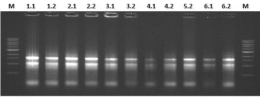
The results of the RNA isolation is shown in figure 4. The 28s rRNA bands appears equal to or more abundant than the 18s rRNA band, thereby indicating that little or no RNA degradation occurred during extraction.
Natural BioBrick
With designed primers, the coat protein gene of the PLRV was isolated and bricked with iGEM prefix and suffix. Because we obtained the biobrick totally from nature and know the potential of VLP coat proteins based on our results with the CCMV and Hepatatis B VLPs, we firmly believe in the significance of the isolated PLRV coat protein. This makes the PLRV coat protein biobrick our favorite natural biobrick.
Results
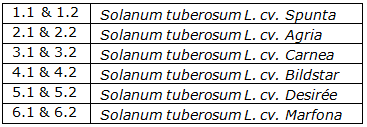
We isolated the PLRV CP genes from multiple potato cultivars (table 1). Four of these were sequenced and the results were used to find natural variations in the viral coat protein: three PLRV coat proteins from different potato cultivars and one PLRV coat protein with a Histidine-tag added to the N-terminal. The three bare PLRV coat proteins were confirmed to be positioned nicely within the iGEM prefix and suffix. Unfortunately the sequence results of the PLRV CP with Histidine-tag showed the absence of the PstI restriction site in the suffix. The three different PLRV coat protein BioBrick parts were submitted to the Registry with the following accession numbers: BBa_K883402, BBa_K88340 and BBa_K883404,
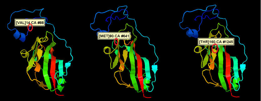
Alignment of the obtained sequences showed 9 single nucleotide polymorphisms (SNP's) of which only 3 resulted in a different amino acid. Amino acid replacement positions can be seen in the figure 5. The PLRV isolated from Solanum tuberosum cv. Desirée (potato cultivar Desirée) showed the substitution of a valine with an alanine, which have very similar hydrophobic properties. Also the two other amino acid variations have more or less similar physical properties. These results show that the structure of the PLRV coat protein is very conserved.
Natural BioBrick
With designed primers, the coat protein gene of the PLRV was isolated and bricked with iGEM prefix and suffix. Because we obtained the biobrick totally from nature and know the potential of VLP coat proteins based on our results with the CCMV and Hepatatis B VLPs, we firmly believe in the significance of the isolated PLRV coat protein. This makes the PLRV coat protein biobrick our favorite natural biobrick.
Read Through part
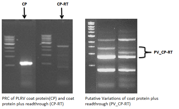
The PLRV read through parts might have a function in the transmission by aphids, which are the vectors which spread the virus. The green peach aphid, Myzus persicae is the most important vector for spreading the virus. We expect that these so-called ‘’spikes’’ can be changed without having a large effect on VLP formation. It is shown that PLRV VLP’s can be formed with and without the read through [4]. We wanted to know to what extent we could modify this spike for application of the plug and apply system (P’NAS). Therefore we isolated the PLRV coat protein including read through part. PCR with primers specific to the start of the coat protein and to the end of the read through part showed DNA segments of various sizes. This might indicate that there is large variation in the size of the read through part. Unfortunately, all attempts to clone and sequence the PLRV rad through part failed.
Isolation of the TuYV constructs
The viral genes encoding the TuYV constructs were obtained from a plasmid encoding the entire viral genome (GenBank: X13063.1). This plasmid was provided to us, via Dr. Kormelink of Wageningen UR’s Virology faculty, by Véronique Brault of the UMR SVQV in Strasbourg. We successfully made four TuYV BioBricks (they are TuYV coat ptotein, TuYV coat protein with His-tag, TuYV coat protein with partial readthrough and TuYV coat protein with partial readthrough with His-tag) from the genome plasmid as well.
References
1. Potato leafroll virus. Available from: http://en.wikipedia.org/wiki/Potato_leafroll_virus.
2. Juergens, M., et al., Genetic analyses of the host-pathogen system Turnip yellows virus (TuYV)-rapeseed (Brassica napus L.) and development of molecular markers for TuYV-resistance. Theor Appl Genet, 2010. 120(4): p. 735-44.
3. Diseases: Potato leafroll luteovirus - Potato Leaf Roll Virus (PLRV) Available from: http://www.agroatlas.ru/en/content/diseases/Solani/Solani_Potato_leafroll_luteovirus/.
4. Lamb, J.W., et al., Assembly of virus-like particles in insect cells infected with a baculovirus containing a modified coat protein gene of potato leafroll luteovirus. J Gen Virol, 1996. 77(Pt 7): p. 1349-58.
 "
"


