Team:Freiburg/Project/Experiments
From 2012.igem.org
(→Activation of transcription) |
|||
| Line 50: | Line 50: | ||
<br> | <br> | ||
| - | == | + | == Direpeat amplification by colony PCR == |
---- | ---- | ||
<br> | <br> | ||
<html> | <html> | ||
| + | <div align="justify">To assess if the direpeats have indeed been successfully cloned into our expression vector, we have accomplished colony PCR with a variety of samples. To this end, we have designed primers which bind to both ends of the direpeat region and thus amplify the direpeats of our TAL protein. | ||
| + | The original vector contains a kill cassette which induces cell killing under certain conditions unless it is replaced by the direpeats during the Golden Gate reaction. | ||
| + | This cassette will also be amplified by the designed primers. A distinction between the amplification of the kill cassette and the direpeats can be made upon the amplicon length: if the kill cassette is amplified, the resulting amplicon will contain 1527bp, while direpeat amplification will produce amplicons of 1276bp length. The difference between these two amplicons is 251bp, and can be detected by agarose gel electrophoresis. (Figure) | ||
| + | |||
| + | <img src="" align="right" width="400px" hspace="20" vspace="20" alt="Direpeat amplification by colony PCR"/> | ||
| + | |||
| + | Indeed, our negative control, consisting of a vector will kill cassette and without direpeats (2nd lane) yields one clear band which lies above those of our positive control, a vector in which the direpeats have been successfully cloned (1st lane). The remaining lanes show the results for our samples of interest. | ||
| + | Obviously, the colony PCR did not produce one single product when amplifying the direpeats, but rather a smear consisting of amplicons with varying lengths. | ||
| + | This is due to numerous homologies within the direpeats. | ||
| + | As it has been described previously, each domain of the TAL protein is homologous to the rest, except for two amino acids which are responsible for the TAL´s specific binding to one of the four DNA nucleotides. These homologies are the reason why PCR amplification of TAL proteins is impossible. | ||
| + | To eliminate those homologies to the greatest extent possible, we changed codon usage within our direpeats. Nevertheless, as the results of our colony PCR demonstrate, there is still a profound amount of homologies left, which imposes difficulties on the amplification of our direpeats (our TAL protein) by PCR and instead results in a smear. | ||
| + | Thus, the existence of this smear indicates the presence of direpeats within the expression vector and therefore points to a successful assembly of a TAL protein. | ||
| + | <br><br><br> | ||
| + | |||
| + | == Activation of transcription == | ||
| + | ---- | ||
| + | <br> | ||
<div align="justify">To show that our TAL effectors are actually working, we used our completed toolkit to produce a TAL protein fused to a VP64 transcription factor. With this TAL-transcription factor construct we targeted a sequence upstream of a minimal promotor coupled to the gene of the enzyme secreted alkaline phosphatse (SEAP). | <div align="justify">To show that our TAL effectors are actually working, we used our completed toolkit to produce a TAL protein fused to a VP64 transcription factor. With this TAL-transcription factor construct we targeted a sequence upstream of a minimal promotor coupled to the gene of the enzyme secreted alkaline phosphatse (SEAP). | ||
Revision as of 20:52, 26 September 2012
Experiments
Gene activation
 This reporter system gives us a couple of advantages over standard egfp or luciferase systems. First of all the SEAP is secreated into the cell culture media, therefore we dont have to destroy our cells to measure, but just take a sample from the supernatant. We are also able to measure one culture multiple times, e.g. at two different time points. Another advantage is the measurement via photometer which makes the samples quantitive compareable.
Experimental design
 The experiment was done with four different transfections, either no plasmid, only the TAL vector, only the SEAP plasmid or a cotransfection of both plasmids. The cells were seeded on a twelve well plate the day before in 500µl culture media per well. The transfection was done with CaCl2 after a cellculture course protocol written by the lab of professor Weber.
Results
The toolkit
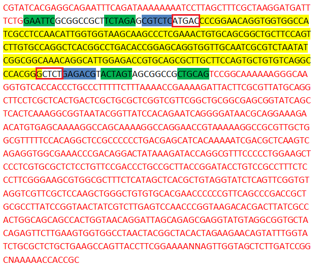
The creation of a toolkit with 96 different parts not only means a lot of labwork but also a lot of organisational tasks, sequencing and analysis. We don't want to bore you with the 96 sequences of our finished biobricks, but we want to give you one example of a finished biobrick and highlight some of the interesting and important strips in its sequence. If you are interested in the other sequences, just have a look at our parts section or go to the Registry of Standard Biological Parts.

|
In this sequence of our biobrick AA1, the main features of all our biobricks are highlighted. As pointed out in the Golden Gate Standard section of our project description, all direpeat plasmids are submitted in the Golden Gate Standard, that was developed by us and which is fully compatible with existing iGEM standards. In yellow you can see the direpeat gene fragment itself, the green parts are iGEM restriction sites (a requirement for all biobricks), the sequence written in red is part of the psb1C3 vector, the blue sequences are recognition sites for BsmB1 and the red boxes are the cutting sites of BsmB1.
Creation of TAL sequences - Golden Gate Cloning
Admittedly, our GATE assembly kit is a little larger than the kit published from the Zhang kit in Nature this year (it comprises 78 parts). But considering that future iGEM teams can easily combine the parts to form more than 67 million different effectors, we believe that it was worth the effort. Now, to get from the toolbox to the finished TAL effector, you only need a few components: six direpeats, one effector backbone plasmid, two enzymes and one buffer. If you mix these components and incubate in your thermocycler for 2.5 hours, you get your custom TAL effector. To put this in perspective: The average turnaround time for TALE construction with conventional kits is about two weeks! In the following sections, we want to show you the results of our trials with the finished biobricks and the sequencing of built TAL effector constructs.
Direpeat amplification by colony PCR
== Activation of transcription == ----
 In theory, the TAL domain should bring the fused VP64 domain in close proximity to the minimal promotor to activate the transcription of the repoter gene SEAP. The phosphatase is secreated an acummulates in the cell culture media. After 24 and 48 hours we took samples from the media, kept them at -20°C, and subjected them two days later to photometric analysis.
As it is observable in the graph, co-transfection of cells with TAL and SEAP plasmids (++) yielded a high increase in SEAP activity, compared to the control samples. The graph shows the average value of three biological replicates with its standard deviation. We further performed a ttest (Table) to prove if our experiment is statistical significant. The yellow highlighted fields show the p-values for our double transfections. As it is clearly observable, the p-values range below a value of 0,05 which indicates that our TAL transcription factor is able to elevate the transcription of the SEAP gene in a statistical significant manner.

After addition of pNPP, the substrate of SEAP, the activity of SEAP was measured over a period of time. In the next image, the results of the first nine minutes of this measurement are shown. After this time, the OD of the double transfection (++) rose too high to be measured by our photometer. As it is clearly visible, the sample with the double transfection shows a profound increase in the OD. This points to the fact that great amounts of SEAP have been secreted into the cell culture media due to elevated gene expression. In the other samples almost no SEAP activity was measureable. The sample transfected with only the SEAP plasmid showed the highest OD but this effect was not statistically significant (p-value:0,25/0,51).
In the samples, that had been taken 48h after double transfection, the same effects could be demonstrated.
Furthermore, we reapeated the same experiment for a second time. The corresponding data can be viewed here:
 "
"

