Contents |
Ligation of the readout into pCEP4 and pCEP4-HA
Protocol: Ligation
Ligation is a method of combining several DNA fragments into a single plasmid. This is often the
step following a PCR (and a PCR cleanup) or a gel extraction. You can also do a "dirty" ligation, where you follow a certain number of digestions directly by a ligation.
- Download the following spreadsheet : File:Team-EPF-Lausanne Ligation.xls
- Fill in the pink areas with the vector and fragment concentration, their size and the ratio.
- Add all the suggested ingredients order in a microcentrifuge tube, in the order they appear.
- Ligate for 2 hours at 14ºC.
- Immediately transform competent bacteria with the ligation product.
Note: This protocol hasn't been optimized for blunt-end ligation (though it might still work).
The readout for the LovTAP experiment was ligated into both versions of its vector at different ratios. The ligations were stored for later transformation.
Chloramphenicol plate preparation
Protocol: Agar Plates
- Add to a bottle:
- 20 g/l of LB broth powder.
- 10 g/l of Agar (not agarose).
- Fill up with DI water and autoclave (program 106, takes 2 hours). Remember to leave the cap loose!
- Label the plates (found in the stock room).
- When the autoclaving is done, close the bottle cap and take the bottle out.
- Let rest until it cools down to around 55ºC (can be held for some seconds): if warmer, the antibiotic degrades, if colder, the broth gelifies.
- Add the antibiotic (Ampicillin 100 µg/ml) = 1 ml of 100 mg/ml Amp for 1l of broth. Same for chloramphenicol or Spe.
- Add 25-30 ml of broth to each plate and let open for 2 hours.
- Close plates, wrap in alu foil (Amp is sensitive to light) and store at +4ºC.
Chloramphenicol plates were prepared for the culture of Biobricks and their empty vector psB1C3 (the antibiotic was at 34 µg/ml, we kept that concentration).
Gel electrophoresis of the colony PCR from the day before
Protocol: Gel Electrophoresis
Agarose concentration depends on the size of the DNA to be run. We will mostly use 1%.
VOL is the desired volume of gel in ml:
CH Lab
- Add 0.01*VOL g of agarose to a clean glass bottle.
- Pour VOL/50 ml of 50xTAE in a graduated cylinder. Fill up to VOL ml with di water.
- Add the resulting VOL ml of 1xTAE to the glass bottle with agarose.
- Microwave, at 7, the bottle (loose cap!) until it boils.
- Carefully remove bottle (can be super heated!) and check for the total absence of particles. Microwave again if needed.
- Prepare a gel box, with comb, and fill it up with the agarose solution (maybe not the whole solution is needed).
- Add 0.05 µl per ml of gel in the box of Red Gel (it's in the iGEM drawer) and stirr until disolved.
- Wait until cold and solidified.
- Carefully remove comb.
- Place the box in the electrophoresis chamber.
- Fill up the electrophresis chamber with 1x TAE buffer.
- Add blue dye to the DNA samples (6x loading buffer, that is 10 µl in 50 µl of DNA solution).
- Inject 30 µl of ladder marker in the first well (that's 1 µg of DNA).
- Inject 60 µl of each DNA solution in the other wells.
- Set voltage to 70-90 V and run for 30-40 min, or until the dye reaches the last 25% of the gel length (DNA travels from - to +).
- Place the gel under the camera, cover, turn UV on and take photos!
Preparing the ladder:
- get 1kb ladder DNA from the freezer (500 µg/ml).
- for 30 charges, 30 µl per charge, we need 900 µl:
- 60 µl of 1kb ladder DNA
- 150 µl of dye (6x loading buffer)
- 690 µl of water
BM Lab
In this lab the gels are slightly different. The total volumes for the small, the medium and the large gel are respectively 60ml, 80ml and 90ml. As we use 0.5x TAE buffer instead of 1x, we can use higher voltages (170V seems to work fine). The gel should run 20-40 minutes, not more. As the gel is thinner, load less DNA (up to ~10ul).
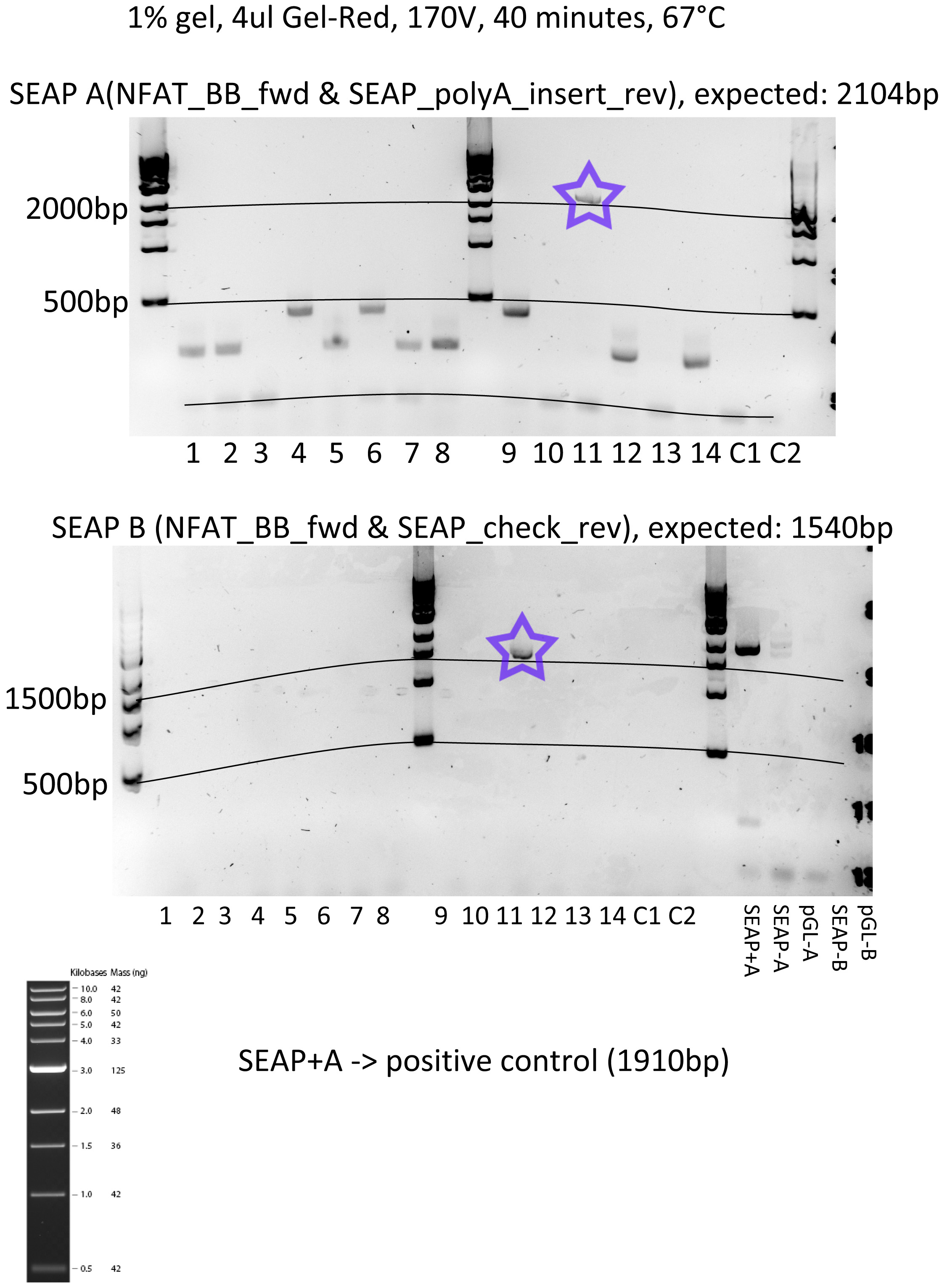
Ran a gel of the pGL-SEAP control PCR with both buffers, found out that only one colony (the 11th one) had worked. Made a glycerol stock out of that one for sequencing and further use.
Plates observation
Took the pHY42 plates out (they were transformed the evening before). Put them at 4°C, to be used for a later maxiprep culture.
 "
"
